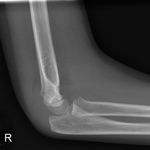
 | This 8 year old girl presented to the Emergency Department following a fall onto her right arm. The fall was unwitnessed. She was examined and found to have a painful swollen right elbow. She was referred for right elbow radiography. The lateral elbow projection image is shown left. |
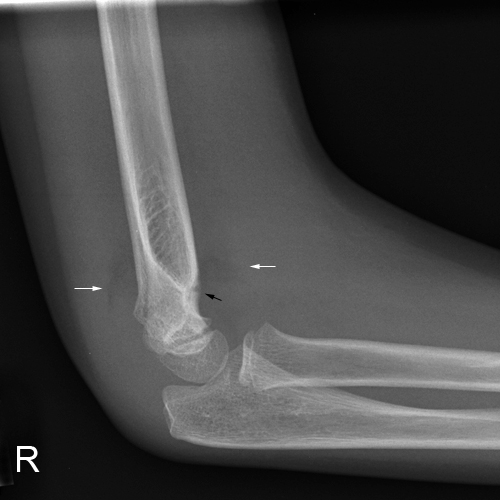
 | There are large anterior and posterior fatpad signs (white arrows) and a defect in the anterior cortex suggesting a supracondylar fracture. |
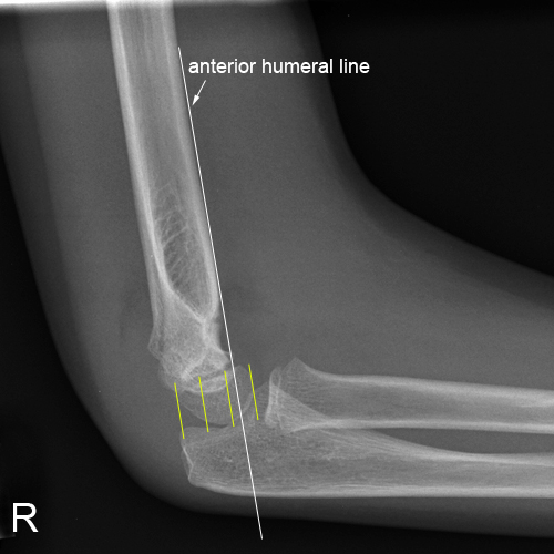
 | The anterior humeral line does not fall into the middle third of the capitellum providing further support for the diagnosis of supracondylar fracture with slight posterior displacement of the angular distal fragment (Gartland type I) |
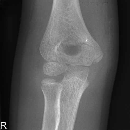
 | The AP elbow projection image demonstrates an intercondylar fracture (white arrow). |


