Elbow Dislocations
Elbow dislocations are reported to be the second most common dislocation behind shoulder dislocations. (Conwell, H.E. 1961 in John Harris et al, The Radiology of Emergency Medicine, 3rd Ed, Williams and Wilkins, 1993, 344). This page examines the radiography of elbow dislocations and associated fractures.
Types of Elbow Dislocations
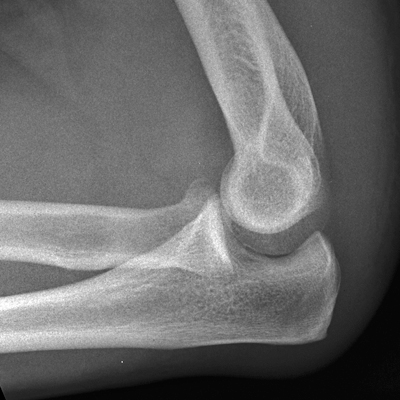
The most common elbow dislocation is a posterior dislocation of the radius and ulna with respect to the distal humerus. Associated fractures may be hidden on the initial plain film imaging and may show only on the post-reduction plain film imaging.
Case 1
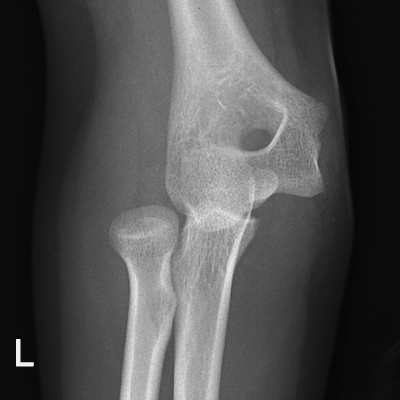
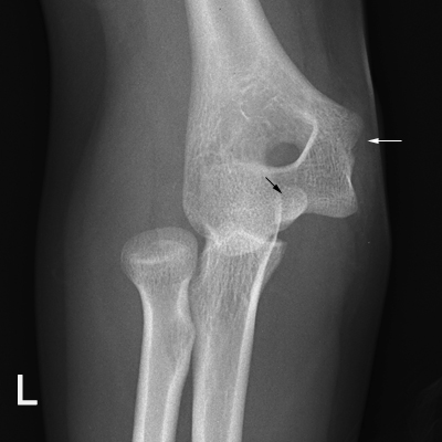
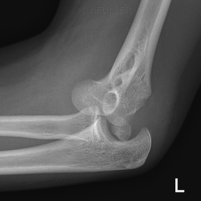
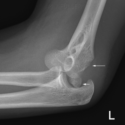
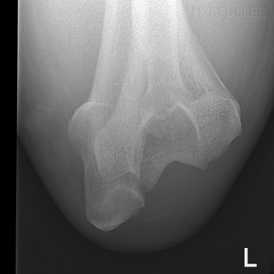
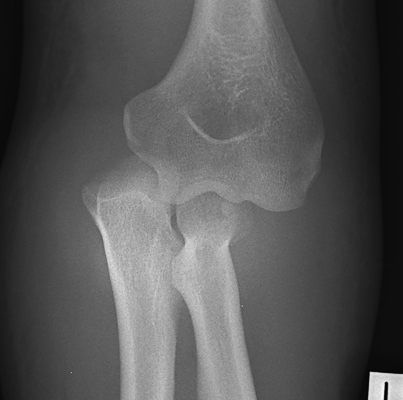
This 14 year old girl fell onto an outstretched left hand and reported that she thought her left elbow had "popped out". The left elbow was painful and deformed. She was referred for elbow radiography.
Case 2
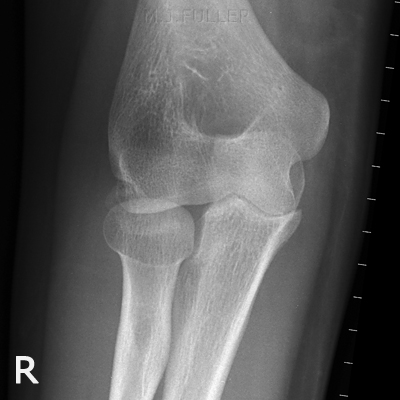
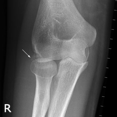
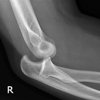
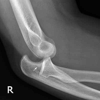
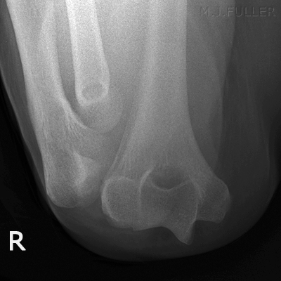
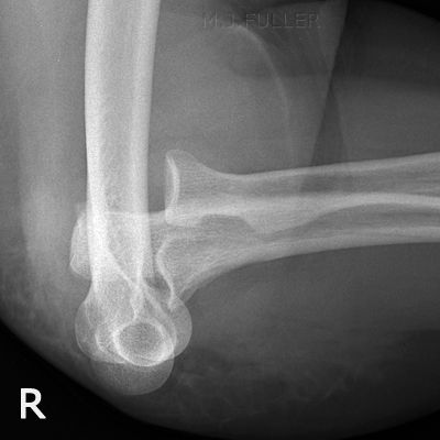
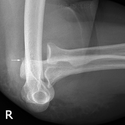
Case 3This 18 year old male presented to the Emergency Department with an unknown history. His right elbow was painful and deformed. He was referred for elbow radiography.
This 70 year old lady presented to the Emergency Department after falling onto her right side. She was examined and found to have a painful and deformed right elbow and was referred for radiography of her right elbow.
Case 4
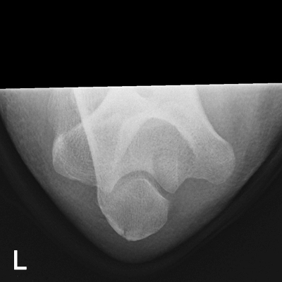
This 34 year old man presented to the Emergency Department after falling from a roof. He was examined and found to have a painful and deformed left elbow and was referred for radiography of his left elbow.
Case 5
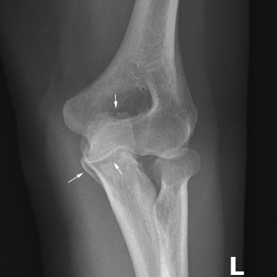
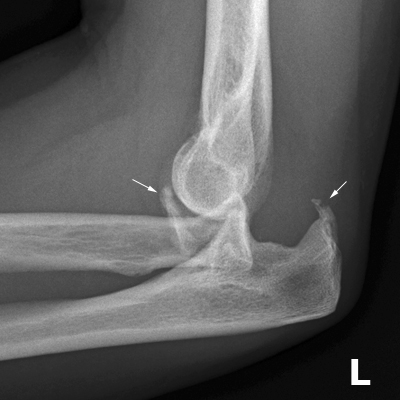
This 57 year old lady presented to the Emergency Department after falling onto her left side. She was examined and found to have a painful and deformed left elbow and was referred for radiography of her left elbow.
Comment
This patient had her elbow dislocation reduced in theatre. On follow-up appointments, the elbow joint was found to be unstable. The patient was referred for CT imaging of her left elbow which revealed multiple fractures including fractures of the coronoid and olecranon. This case demonstrates the common finding that posterior dislocations of the elbow are often accompanied by fractures. There is a danger of satisfaction syndrome when the initial imaging is performed- the radiographer should be aware of the potential for concomittant fractures and ensure that every effort is made to account for the source of the bony fragments demonstrated.
...back to the Wikiradiography home page
...back to the Applied Radiography home page
