Imaging Radial Head Fractures

From wikiRadiography
Jump to navigation Jump to search

Introduction

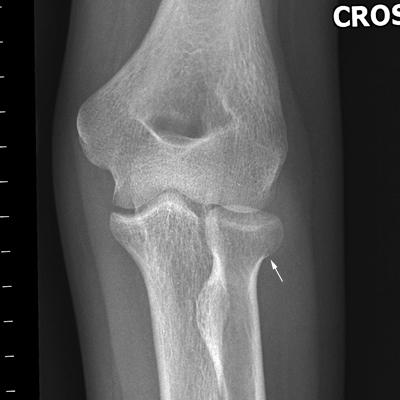
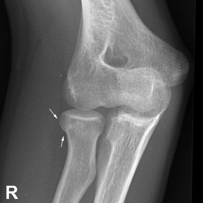
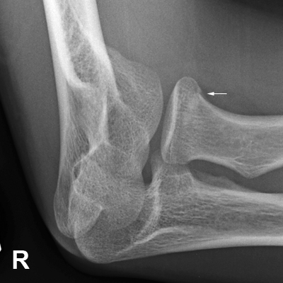
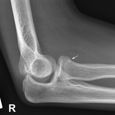
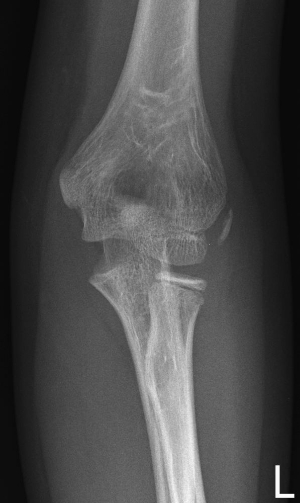
Radial head and/or neck fracture is a commonly associated with a fall onto an outstretched hand in adults. This page considers radial head radiographic technique and pathological appearances.


Anatomy

Ossification

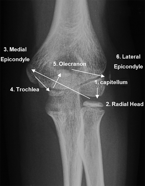
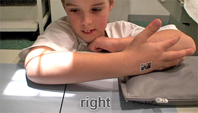
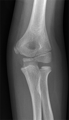
ossification of the elbowThe paediatric elbow can present a confusing array of ossifications. The ossification centres generally ossify in the order shown (left).

A useful mnemonic for remembering the order of the ossification centres is the CRITOL rule

Capitellum
R
adial head
Internal (medial epicondyle)
T
rochlea
O
lecranon
L
ateral epicondyle

It is noteworthy that the ossification centres of the elbow do not always follow this order of ossification.
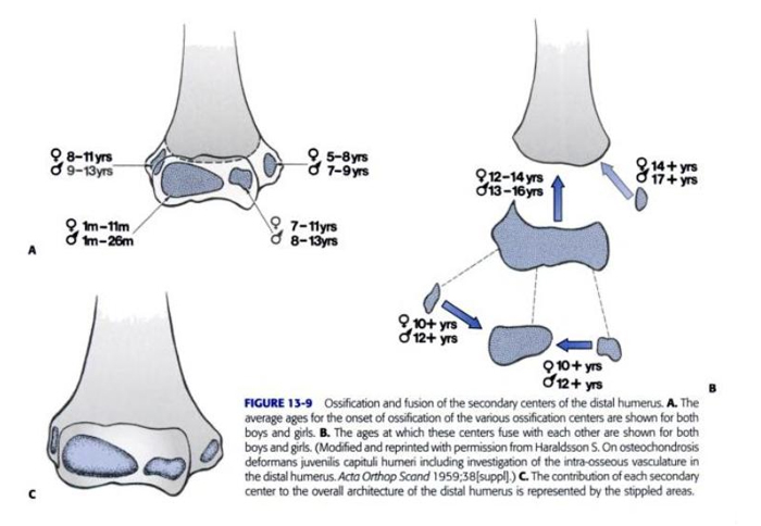
ossification of the elbow


<a class="external" href="http://books.google.com.au/books?id=6AXkE4y1678C&pg=PA509&lpg=PA509&dq=radial+head+fractures+incidence+in+adults&source=bl&ots=JRFuoZ23Kh&sig=r6wMfpmEAfwn6_ySqvaTdogcJVk&hl=en&ei=u_FHSvmRIIfU7AOqkZnoAw&sa=X&oi=book_result&ct=result&resnum=9" rel="nofollow" target="_blank">Charles A. Rockwood, Kaye E. Wilkins, James H. Beaty, James R. Kasser
Rockwood and Wilkins' fractures in children
Lippincott Williams & Wilkins, 2006</a>
It can also be helpful in distinguishing fractures from normal ossification centres to be aware of the ages of appearance and fusion of the elbow epiphyses and apophyses.


Elbow Joint Effusion

elbow joint effusion
<a class="external" href="http://books.google.com.au/books?id=6AXkE4y1678C&pg=PA509&lpg=PA509&dq=radial+head+fractures+incidence+in+adults&source=bl&ots=JRFuoZ23Kh&sig=r6wMfpmEAfwn6_ySqvaTdogcJVk&hl=en&ei=u_FHSvmRIIfU7AOqkZnoAw&sa=X&oi=book_result&ct=result&resnum=9" rel="nofollow" target="_blank">adapted from the Journal of Bone and Joint Surgery in</a>
<a class="external" href="http://books.google.com.au/books?id=6AXkE4y1678C&pg=PA509&lpg=PA509&dq=radial+head+fractures+incidence+in+adults&source=bl&ots=JRFuoZ23Kh&sig=r6wMfpmEAfwn6_ySqvaTdogcJVk&hl=en&ei=u_FHSvmRIIfU7AOqkZnoAw&sa=X&oi=book_result&ct=result&resnum=9" rel="nofollow" target="_blank">Charles A. Rockwood, Kaye E. Wilkins, James H. Beaty, James R. Kasser</a>
<a class="external" href="http://books.google.com.au/books?id=6AXkE4y1678C&pg=PA509&lpg=PA509&dq=radial+head+fractures+incidence+in+adults&source=bl&ots=JRFuoZ23Kh&sig=r6wMfpmEAfwn6_ySqvaTdogcJVk&hl=en&ei=u_FHSvmRIIfU7AOqkZnoAw&sa=X&oi=book_result&ct=result&resnum=9" rel="nofollow" target="_blank">Rockwood and Wilkins' fractures in children</a>
<a class="external" href="http://books.google.com.au/books?id=6AXkE4y1678C&pg=PA509&lpg=PA509&dq=radial+head+fractures+incidence+in+adults&source=bl&ots=JRFuoZ23Kh&sig=r6wMfpmEAfwn6_ySqvaTdogcJVk&hl=en&ei=u_FHSvmRIIfU7AOqkZnoAw&sa=X&oi=book_result&ct=result&resnum=9" rel="nofollow" target="_blank">Lippincott Williams & Wilkins, 2006</a>
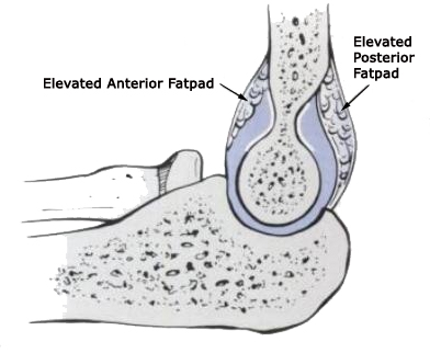
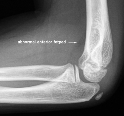
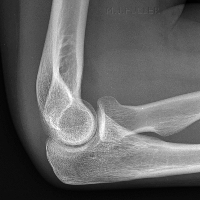
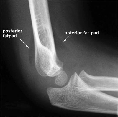
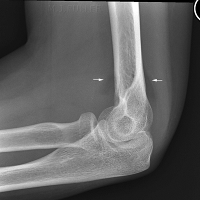
The elbow joint is a synovial joint. When an elbow injury occurs and there is an intra-articular involvement, there can be an associated elbow effusion. As the elbow joint distends with fluid, the adjacent soft tissues can be displaced. The anterior and posterior fat pads are of particular interest to radiographers because they can provide a valuable indicator of intra-articular injury when the elbow fat pads are displaced by an elbow joint effusion. Importantly, evidence of an elbow joint effusion on elbow plain film images does not indicate a definite elbow fracture- it does indicate an elbow joint injury.

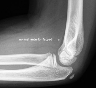
normal anterior elbow fatpad
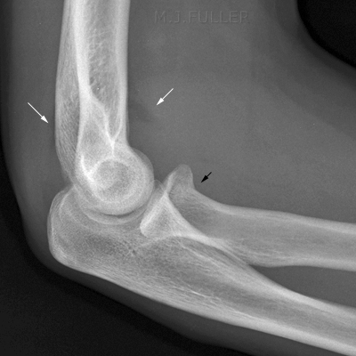
The anterior elbow fat pad can often be seen in a normal lateral elbow as a stripe of radiolucency parallel to the anterior cortex of the distal humerus. A visible anterior fat pad can be a normal finding- a visible posterior fatpad is not a normal finding.
abnormal anterior elbow fatpadelbow joint effusion
<a class="external" href="http://books.google.com.au/books?id=6AXkE4y1678C&pg=PA509&lpg=PA509&dq=radial+head+fractures+incidence+in+adults&source=bl&ots=JRFuoZ23Kh&sig=r6wMfpmEAfwn6_ySqvaTdogcJVk&hl=en&ei=u_FHSvmRIIfU7AOqkZnoAw&sa=X&oi=book_result&ct=result&resnum=9" rel="nofollow" target="_blank">adapted from the Journal of Bone and Joint Surgery in</a>
<a class="external" href="http://books.google.com.au/books?id=6AXkE4y1678C&pg=PA509&lpg=PA509&dq=radial+head+fractures+incidence+in+adults&source=bl&ots=JRFuoZ23Kh&sig=r6wMfpmEAfwn6_ySqvaTdogcJVk&hl=en&ei=u_FHSvmRIIfU7AOqkZnoAw&sa=X&oi=book_result&ct=result&resnum=9" rel="nofollow" target="_blank">Charles A. Rockwood, Kaye E. Wilkins, James H. Beaty, James R. Kasser</a>
<a class="external" href="http://books.google.com.au/books?id=6AXkE4y1678C&pg=PA509&lpg=PA509&dq=radial+head+fractures+incidence+in+adults&source=bl&ots=JRFuoZ23Kh&sig=r6wMfpmEAfwn6_ySqvaTdogcJVk&hl=en&ei=u_FHSvmRIIfU7AOqkZnoAw&sa=X&oi=book_result&ct=result&resnum=9" rel="nofollow" target="_blank">Rockwood and Wilkins' fractures in children</a>
<a class="external" href="http://books.google.com.au/books?id=6AXkE4y1678C&pg=PA509&lpg=PA509&dq=radial+head+fractures+incidence+in+adults&source=bl&ots=JRFuoZ23Kh&sig=r6wMfpmEAfwn6_ySqvaTdogcJVk&hl=en&ei=u_FHSvmRIIfU7AOqkZnoAw&sa=X&oi=book_result&ct=result&resnum=9" rel="nofollow" target="_blank">Lippincott Williams & Wilkins, 2006</a>


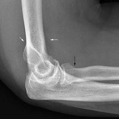
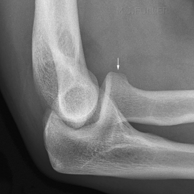
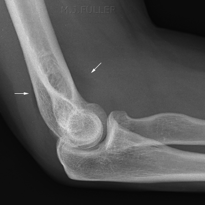
In the presence of an elbow effusion, the fatpads can be displaced in an appearance that is commonly referred to as a sail sign. When you see a sail sign on a lateral elbow image, consider carefully whether you have adequately demonstrated a related fracture, and if not, whether supplementary views would be worthwhile to determine if a fracture exists. Note also a subtle posterior fatpad sign (left)- this is subtle but important in that it suggests a likelihood of fracture at about the 90% level.
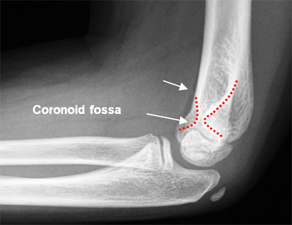
lateral elbowThe distal humerus has two fossae- the coronoid fossa accommodates the coronoid process of the ulna on elbow flexion and the olecranon fossa accommodates the olecranon process of the ulna on elbow extension. The coronoid fossa is a shallower fossa than the olecranon fossa- this is why the anterior fat pad can appear as a normal elbow feature and also explains why an anterior fat pad sign can appear without the posterior fat pad sign. i.e. it takes a larger elbow effusion to displace the posterior fat pad out of the deeper olecranon fossa than the anterior fat pad out of the shallower coronoid fossa.

Lateral Elbow Attempt 1
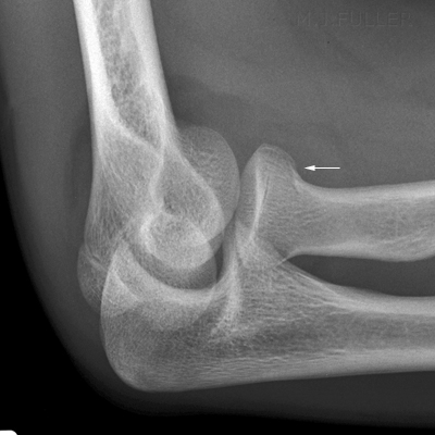
LATERAL ELBOW
Lateral Elbow Attempt 2
LATERAL ELBOW
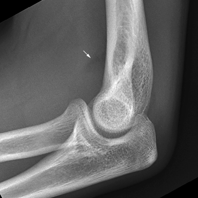
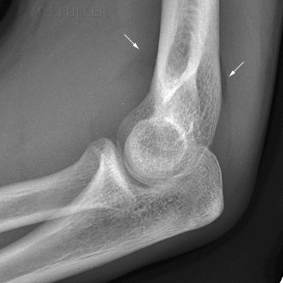
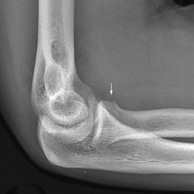
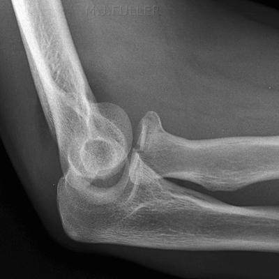
This is a lateral elbow image of a patient with an intra-articular injury. There is a clearly demonstrated anterior fatpad sign (arrowed) and a suggestion of a posterior fatpad sign.The repeat lateral elbow image, whilst failing to achieve a true lateral position, achieved an improved demonstration of the anterior and posterior fatpads (note that occasionally a lateral elbow image will demonstrate the distended joint- this is such a case).


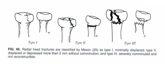
Mason's Classification of Radial Head Fractures
classificatiopn of radial head fractures
<a class="external" href="http://books.google.com.au/books?id=iJZyb0s7dhgC&pg=RA3-PA341&lpg=RA3-PA341&dq=elbow+radiography&source=bl&ots=VlMjhi5fob&sig=yGvshuYLxyz9yaOj4z-51_NBV6w&hl=en&ei=A3VJSon3LZWVkAWX_sjvCQ&sa=X&oi=book_result&ct=result&resnum=5" rel="nofollow" target="_blank">The Radiology of Emergency Medicine, </a>
<a class="external" href="http://books.google.com.au/books?id=iJZyb0s7dhgC&pg=RA3-PA341&lpg=RA3-PA341&dq=elbow+radiography&source=bl&ots=VlMjhi5fob&sig=yGvshuYLxyz9yaOj4z-51_NBV6w&hl=en&ei=A3VJSon3LZWVkAWX_sjvCQ&sa=X&oi=book_result&ct=result&resnum=5" rel="nofollow" target="_blank">4th Edition John H. Harris, Jr and William H. Harris (Eds), </a>
<a class="external" href="http://books.google.com.au/books?id=iJZyb0s7dhgC&pg=RA3-PA341&lpg=RA3-PA341&dq=elbow+radiography&source=bl&ots=VlMjhi5fob&sig=yGvshuYLxyz9yaOj4z-51_NBV6w&hl=en&ei=A3VJSon3LZWVkAWX_sjvCQ&sa=X&oi=book_result&ct=result&resnum=5" rel="nofollow" target="_blank">Lippincott, Williams & Wilkins, Philadelphia, 2000</a>
Mason's classified of radial head/radial neck fractures according to the degree of displacement and comminution.


Radiographic Technique

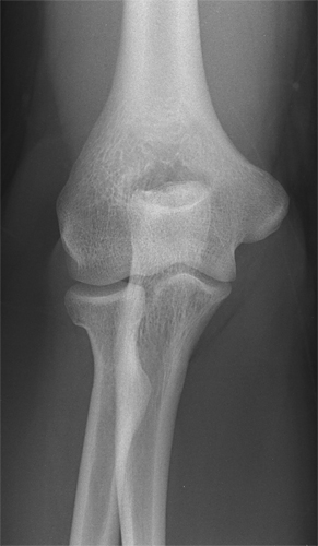
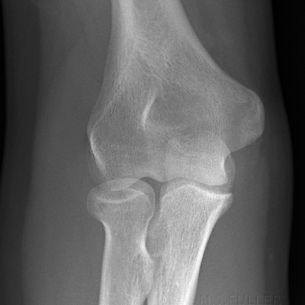
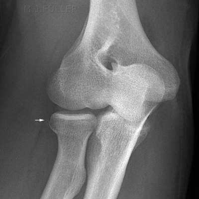
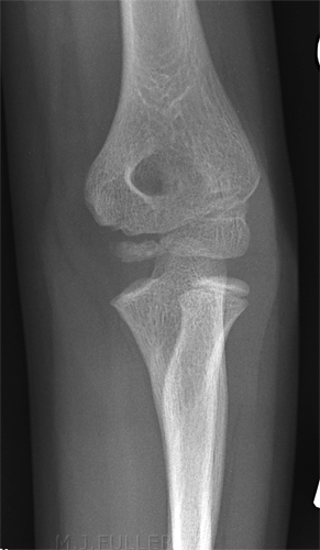
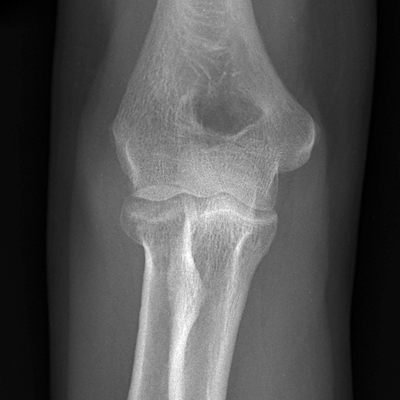
AP Elbow

Option1Option2Option3Option4
AP elbowAP elbowAP elbowAP ELBOW
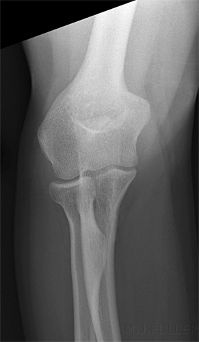
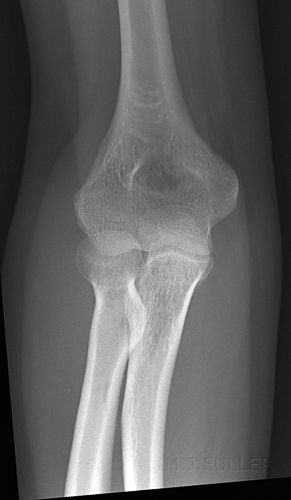
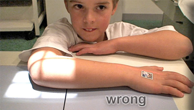
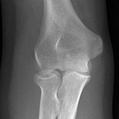
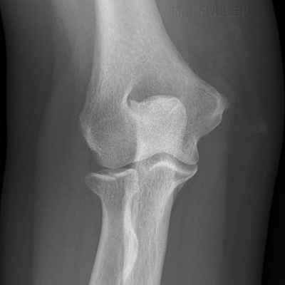
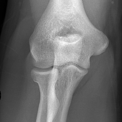
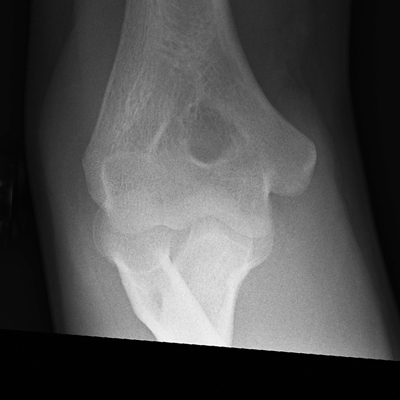
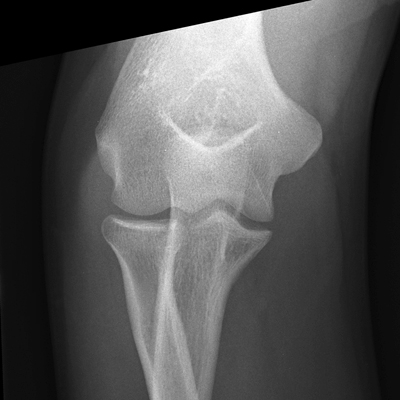
Conventional AP elbow positionThis patient was unable/unwilling to straighten/extend his/her elbow. The image was taken with the forearm resting on the IR. Note the distorted distal humerus anatomy.This patient was unable/unwilling to straighten/extend his/her elbow. The image was taken with the humerus resting on the IR. Note the distorted radius- the radial head is not demonstrated in profile.

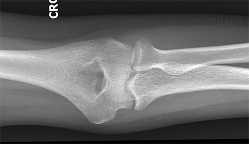
One of the interesting aspects of this view is that it can be the only view that demonstrates a radial head fracture. This may be due to the fact that you achieve a partial enface view that is not achieved in other projections.
This is a compromise technique employed when the patient has a fixed flexion of the elbow and the radiographer is unsure if the bony injury is to the humerus or forearm.


AP ELBOWAP ELBOW
This is an AP view of a partially flexed elbow with the forearm in contact with the image receptor. Note that the radial head is demonstrated in profile. The appearance of the radial head is suggestive of a fracture. This is the same patient with the partially flexed elbow in an AP position with the upper arm in contact with the image receptor. This produces a partially enface view of the radial head providing a clearer demonstration of the comminuted fracture. This considered a compromise view of the elbow but can be utilised to advantage with some types of radial head fracture.


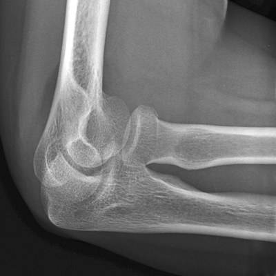
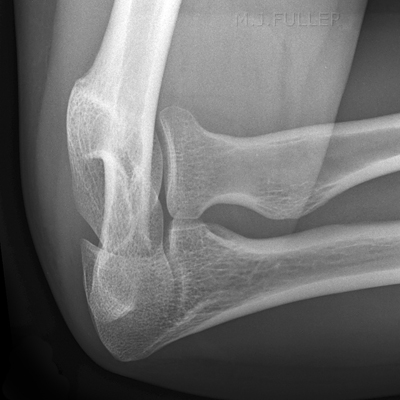
Lateral Elbow

LATERAL ELBOW
<a class="external" href="http://www.radiologyassistant.nl/en/4214416a75d87" rel="nofollow" target="_blank">www.radiologyassistant.nl/en/4214416a75d87</a>
LATERAL ELBOW
LATERAL ELBOW
<a class="external" href="http://www.radiologyassistant.nl/en/4214416a75d87" rel="nofollow" target="_blank">www.radiologyassistant.nl/en/4214416a75d87</a>


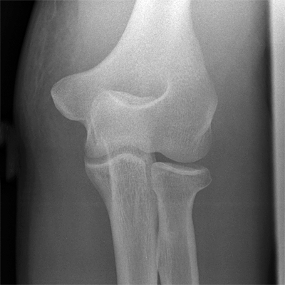
Oblique Elbow

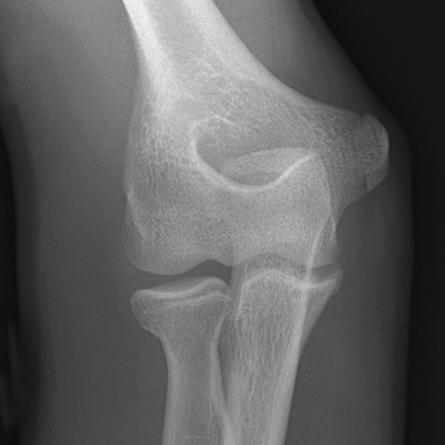
OBLIQUE ELBOWoblique elbow
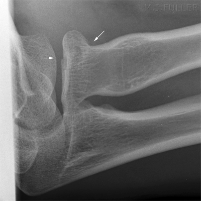
The conventional oblique view of the elbow is employed to demonstrate the radial head. This position projects the radial head off the ulna.This oblique view of the elbow was achieved with angulation of the x-ray tube across the AP elbow rather than with external rotation of the patient's elbow. The image demonstrates some elongation/distortion of the anatomy- this is nevertheless an effective alternative to the conventional oblique elbow technique.


Reverse Oblique Elbow

AP Elbow
AP elbow
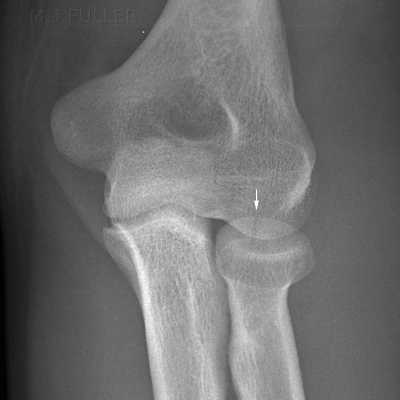
Reverse Oblique Elbow
REVERSE OBLIQUE
The conventional AP elbowThe reverse oblique elbow position requires the internal rotation of the forearm rather than the conventional external rotation. Whilst this position superimposes the radial head over the ulna, radial head fractures can be demonstrated that will not be seen in the conventional views.


The radial head, capitellum view

radial head view posoitioning
<a class="external" href="http://books.google.com.au/books?id=iJZyb0s7dhgC&pg=RA3-PA341&lpg=RA3-PA341&dq=elbow+radiography&source=bl&ots=VlMjhi5fob&sig=yGvshuYLxyz9yaOj4z-51_NBV6w&hl=en&ei=A3VJSon3LZWVkAWX_sjvCQ&sa=X&oi=book_result&ct=result&resnum=5" rel="nofollow" target="_blank">The Radiology of Emergency Medicine, </a>
<a class="external" href="http://books.google.com.au/books?id=iJZyb0s7dhgC&pg=RA3-PA341&lpg=RA3-PA341&dq=elbow+radiography&source=bl&ots=VlMjhi5fob&sig=yGvshuYLxyz9yaOj4z-51_NBV6w&hl=en&ei=A3VJSon3LZWVkAWX_sjvCQ&sa=X&oi=book_result&ct=result&resnum=5" rel="nofollow" target="_blank">4th Edition John H. Harris, Jr and William H. Harris (Eds), </a>
<a class="external" href="http://books.google.com.au/books?id=iJZyb0s7dhgC&pg=RA3-PA341&lpg=RA3-PA341&dq=elbow+radiography&source=bl&ots=VlMjhi5fob&sig=yGvshuYLxyz9yaOj4z-51_NBV6w&hl=en&ei=A3VJSon3LZWVkAWX_sjvCQ&sa=X&oi=book_result&ct=result&resnum=5" rel="nofollow" target="_blank">Lippincott, Williams & Wilkins, Philadelphia, 2000</a>
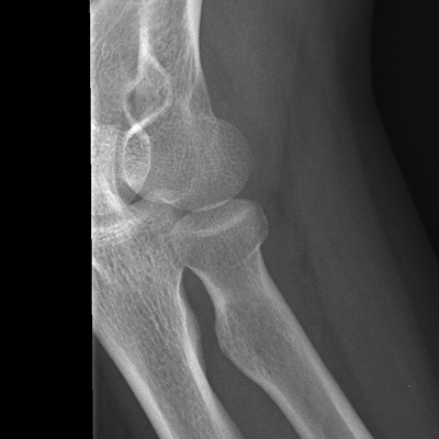
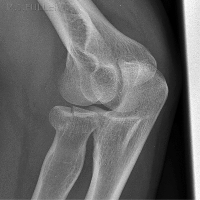
The radial head view is often the only projection that demonstrates a subtle radial head fracture. To perform this view correctly the following are noteworthy
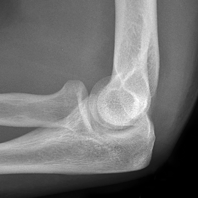
  • the elbow should be in a true lateral position. This is often impossible in a patient with a radial head fracture. In particular, rotation of the hand into a lateral position may be difficult.
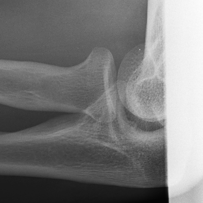
  • a 45 degree tube angle will produce an elongated view of the anatomy but will be more likely to project the radial head clear of the ulna
  • learn where the radial head is positioned in this view and collimate the X-ray beam to include the anatomy of interest only.

further reading
Greenspan A, Norman A. The radial head, capitellum view: useful technique in elbow trauma. AJR Am J Roentgenol. 1982 Jun;138(6):1186-8.


30 Degree Tube Angle
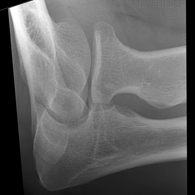
RADIAL HEAD VIEW
45 Degree Tube Angle
RADIAL HEAD VIEW
Some radiographers prefer a 30 degree tube angle for radial head views. With a 45 degree tube angulation and the elbow in a true lateral position you should produce an image of the radial head as shown above. Note that the radial head is largely projected clear of the ulna.


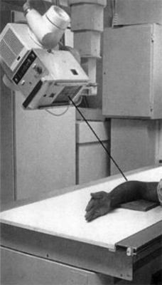
Horizontal Ray Elbow Radiography

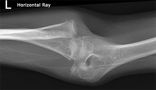
AP elbowThis does not appear to be substantially different to a conventional AP elbow image. It does differ from a conventional AP elbow in the technique employed- this is a horizontal ray technique employed in a patient who was unable/unwilling to move their arm. The image has been presented on the side to emphasize the technique involved. You would arguably not present the image in this orientation for reporting.


Imaging Radial Head Fractures - wikiRadiographyAnother horizontal ray AP elbow. This horizontal ray AP elbow view image demonstrates a common problem with this view- artifact. If possible, support the elbow on a non-opaque material


Image Interpretation

The Fat Pad sign

elbow fatpads
  • Indicates intra-articular injury

  • If the anterior fat pad is visible, 75% chance of articular fracture

  • If the posterior fat pad is visible, 90% chance of articular fracture

  • may not be seen on poor lateral elbow radiograph

  • may not be seen if capsule is ruptured

  • Anterior fat-pad is commonly visible in normal lateral elbow


Case Studies

Case 1

This 20 year old male presented to the Emergency Department following a fall from his skateboard. He had pain in his right elbow and a limited range of movement. He was referred for elbow radiography.

AP ELBOWThe patient was experiencing difficulty extending his elbow. The radiographer performed a compromise flexed AP elbow position which neither favoured demonstration of the forearm nor humerus. The radial head demonstrates an unexplained horizontal sclerotic linear opacity as well as several subtle suspect vertical lucent lines. The appearances are not conclusively abnormal.
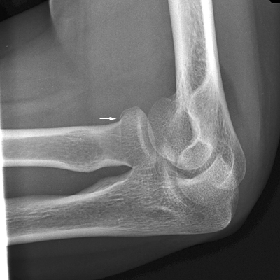
LATERAL ELBOWThere is an anterior fatpad sign (top white arrow)

The supinator fatpad is not clearly abnormal

There is evidence of a radial head fracture (short white arrow)

Note that there is possibly also a posterior fat pad sign that is not demonstrated on this lateral elbow due to malpositioning
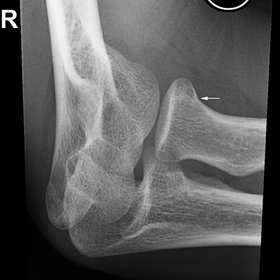
RADIAL HEAD VIEWThe 45 degree radial head view image demonstrates the radial head fracture conclusively.

This is a type I fracture using the Mason classification (i.e. <2mm displacement)

Comment

The radial head fracture is most convincingly demonstrated on the 45 degree radial head view. This is often the case with radial head fractures.


Case 2

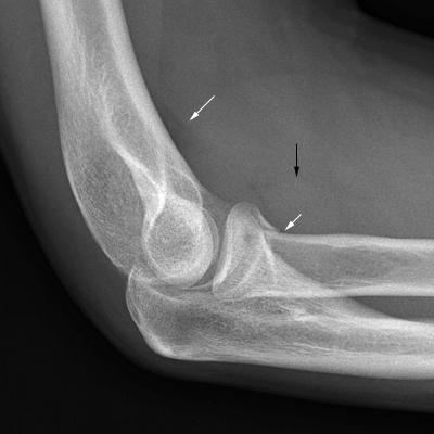
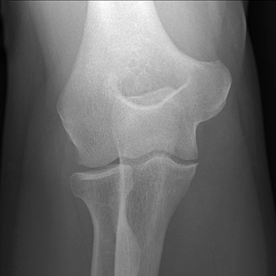
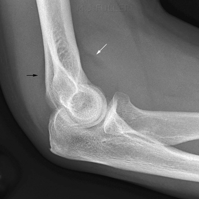
This 38 year old male presented to the Emergency Department following a fall onto an outstretched hand . He had pain in his right elbow and a limited range of movement. He was referred for elbow radiography.

AP ELBOWThe AP elbow view demonstrates the patient has achieved a full extended elbow position. There is no displaced fracture demonstrated
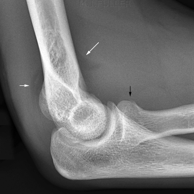
LATERAL ELBOWThis is a good lateral elbow position


There is an anterior fatpad sign ( white arrow)

There is a posterior fatpad sign (black arrow)

The supinator fat pad is not well seen

There is no displaced fracture demonstrated
RADIAL HEAD VIEWThe mechanism of injury and the fatpad signs demonstrated on the routine views warranted further imaging

The 45 degree radial head view image demonstrates no radial head fracture conclusively.

Comment

It is possible that this patient has not sustained any bony injury. It is equally possible that the patient has an undisplaced radial head fracture that has not been demonstrated on this examination.


Case 3

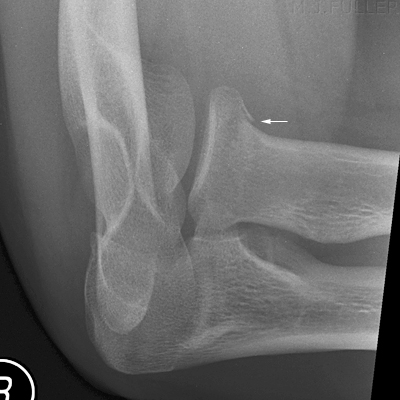
This 15 year old male presented to the Emergency Department following a fall onto an outstretched hand after falling off his pushbike. He had pain in his right elbow and a decreased range of movement. He was referred for elbow radiography.

AP ELBOWThe AP elbow position appears to be over-rolled into an oblique position. This demonstrates the radial head well but was not the intended position.

The bony anatomy is unremarkable
LATERAL ELBOWThis is a slightly malpositioned lateral elbow view.

There is an anterior fatpad sign ( longer white arrow)

There is a posterior fatpad sign (short white arrow)

The supinator fat pad is not well seen

There is discontinuity of the contour of the radial head suggesting a radial head fracture
RADIAL HEAD VIEWThe mechanism of injury and the fatpad signs demonstrated on the routine views warranted further imaging

The 45 degree radial head view image demonstrated a similar discontinuity of the radial head

It is likely that the patient had a type I radial head fracture


Case 4

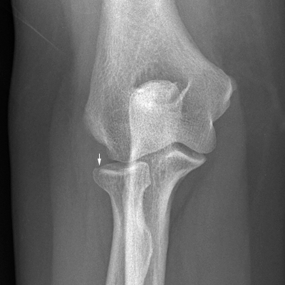
This 56 year old lady presented to the Emergency Department following a fall from steps onto an outstretched hand . She had pain in her right elbow and a decreased range of movement. She was referred for elbow radiography.

AP ELBOWThe AP elbow position appears to be slightly under-rolled into a partly reverse oblique position. This tends to superimpose the medial aspect of the radius over the olecranon.

There is evidence of a radial head fracture (arrowed)
oblique elbowThis is an attempted oblique elbow position that appears to be closer to an AP elbow position.

The radial head fracture is clearly demonstrated
LATERAL ELBOWThis is an attempted lateral elbow view. The humerus is not at right angles to the beam resulting in a malpositioned lateral elbow view.

The anterior and posterior fatpad signs are probably present but not well demonstrated on the malpositioned view.

There is clear evidence of a type I radial head fracture.


Case 5

This 17 year old girl presented to the Emergency Department after falling from a car bonnet . She had pain in her left elbow and a decreased range of movement. She was referred for elbow radiography.

AP elbowThe AP elbow was performed using a cross-table technique. There is a suggestion of a lucent line in a vertical/oblique orientation through the radial head.
LATERAL ELBOWThe lateral elbow position is not a true lateral. Despite this limitation, there are clear anterior and posterior fatpad signs (arrowed).
OBLIQUE ELBOWThis is an attempted oblique elbow view. The elbow has been malpositioned in the collimated field resulting in non-demonstration of the medial elbow anatomy. The radial head has been successfully demonstrated and no displaced fracture is evident.
RADIAL HEAD VIEWThis is a 30 degree tube angle radial head view demonstrating a Mason type I radial head fracture.

Comment

Subtle radial head fractures may only be demonstrated conclusively on the 45 degree radial head view image.


Case 6

This 30 year old man presented to the Emergency Department after falling during a game of soccer . He had pain and swelling in his right elbow and a decreased range of movement. He was referred for elbow radiography.

AP elbowThe AP elbow view image is unremarkable. (the elbow is slightly flexed in this view)
LATERAL ELBOWThe lateral elbow position is not a true lateral. Despite this limitation, there is a subtle cortical disruption of the radial head suggesting the possibility of a radial head fracture. In addition, an anterior fatpad sign is visible and possibly a very subtle posterior fatpad sign.
RADIAL HEAD VIEWThis is a malpositioned lateral view image demonstrating a Mason type I radial head fracture.
OBLIQUE ELBOWThe oblique view of the radial elbow similarly demonstrates a subtle cortical defect in the radial head.


Case 7

This 19 year old male presented to the Emergency Department after falling off his pushbike. He had pain and a decreased range of movement in his right elbow. He was unable to pronate and supinate his right hand. He was referred for elbow radiography.

AP ELBOWThis is an AP elbow view image with the elbow in partial flexion with the patient's hand pronated (note the radius crossing the ulna). The positioning is biased to demonstrate the humerus. (see option 3 above)

No displaced fracture is clearly demonstrated.
AP ELBOWThis is also an AP elbow view image with the forearm in contact with the image receptor. Note that the radius crosses the ulna indicating that the wrist is probably pronated instead of supinated.

No displaced fracture is clearly demonstrated.
LATERAL ELBOWThe lateral elbow view image demonstrates anterior and posterior fatpad signs (white arrows). The radial head appearance is suspicious of a radial head fracture (black arrow)
RADIAL HEAD VIEWThis is a 30 degree radial head view image. There is a clearly demonstrated fracture of the radial head (Mason type II)

Comment

The improved demonstration of the radial head fracture on the dedicated radial head view illustrates the value of this projection


Case 8

This 23 year old male presented to the Emergency Department after falling off his skateboard. He had an extremely swollen and painful left elbow. He was referred for elbow radiography.

AP ELBOWThis is also an AP elbow view image with the elbow in partial flexion and the forearm in contact with the image receptor. There are several lucent lines through the radial head suggesting a comminuted fracture.
AP ELBOWThis is an AP elbow view image with the elbow in partial flexion. The positioning is biased to demonstrate the humerus. (see option 3 above)


There is a fracture of the radial head- possibly comminuted in a peace sign configuration (also called a Mercedes sign)

mercedes signpeace sign<a class="external" href="http://eorif.com/Elbowforearm/Radial+Head+fx.html" rel="nofollow" target="_blank">radial head fracture</a>
<a class="external" href="http://eorif.com/Elbowforearm/Radial+Head+fx.html" rel="nofollow" target="_blank">http://eorif.com/Elbowforearm/Radial%20Head%20fx.html</a>
LATERAL ELBOWThe lateral elbow view image demonstrates anterior and posterior fatpad signs- they are not sharply delineated but, nevertheless, are strongly suggested.
RADIAL HEAD VIEWThis is a 30 degree radial head view image. No displaced fracture is convincingly demonstrated.
REVERSE OBLIQUE ELBOWThis is a reverse oblique view image. A vertical fracture line through the radial head is demonstrated.

Comment

This case demonstrates the value of the AP elbow view with the patient's elbow in partial flexion and the humerus in contact with the image receptor. This view produces a partially axial projection of the radial head which demonstrated the nature of this fracture well.


Case 9

This 16 year old girl presented to the Emergency Department after falling onto her left arm. She had a painful left elbow. She was referred for elbow radiography.

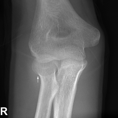
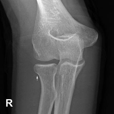
AP ELBOWThis is a modified AP elbow position with the upper arm/humerus in contact with the IR and the elbow in partial flexion. There is a suggestion of a radial head cortical irregularity (arrowed).
AP ELBOWThis is the matching AP view of the flexed elbow with the forearm in contact with the IR. Note that the radial head is demonstrated in true profile compared with the AP view image above. There is a cortical discontinuity of the radial head (arrowed).
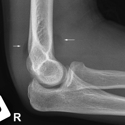
LATERAL ELBOWThe lateral elbow view image clearly demonstrates an anterior fatpad sign (arrowed) with a less obvious posterior fat pad sign (unmarked).
LATERAL ELBOWThe lateral view was repeated which resulted in an improved demonstration of the posterior fatpad.
AP ELBOWIn desperation to clearly demonstrate the suspected fracture of the radial head, the radiographer performed an AP view of the radial head with tube angulation toward the elbow. This is essentially the same as the initial AP elbow projection with tube angulation in lieu of elbow flexion. The radial head fracture is similarly demonstrated (white arrow).

Comment

This case demonstrates the subtlety of some radial head fractures. Undisplaced fractures of the radial head are sometimes impossible to demonstrate on first presentation and may not show until the second, or even third radiographic examination. Note also that subtle differences in the lateral elbow position can improve the demonstration of the elbow fat pads.


Case 10

This 43 year old lady presented to the Emergency Department after falling onto an outstretched hand. She had a painful left elbow which she was unable to extend. She was referred for elbow radiography.

AP ELBOWThe AP elbow view demonstrates partial elbow flexion. There are several indistinct lucent lines through the radial head suspicious of a fracture.
LATERAL ELBOWThere are indistinct, but nevertheless convincing, anterior and posterior fatpad signs (arrowed).
RADIAL HEAD VIEWThe radial head view demonstrates a minimally displaced fracture of the radial head
OBLIQUE ELBOWThe oblique view of the elbow demonstrates the radial head fracture convincingly.

Comment

This case demonstrates the potential value of the external oblique view of the elbow in demonstrating radial head fractures.


Case 11

This 15 year old male presented to the Emergency Department after falling onto his right arm. His elbow was examined and found to be swollen and there was a limited range of movement in the elbow joint. He was referred for right elbow radiography.

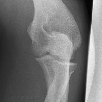
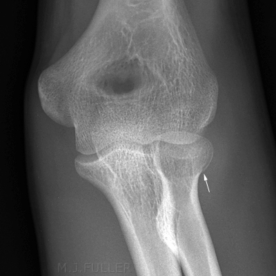
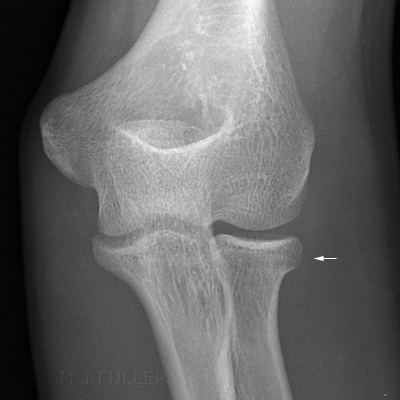
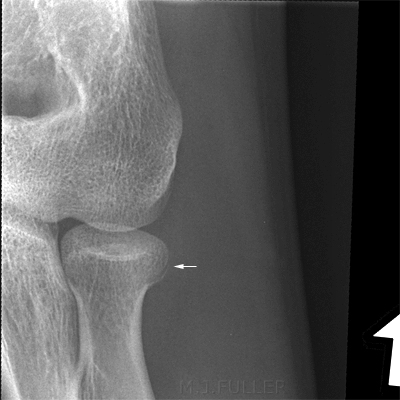
ap elbowThe patient was unable/unwilling to extend his elbow. This AP elbow view image is taken with the upper arm in contact with the IR. This causes distortion of the bony forearm anatomy. There is a very subtle step/discontinuity of the radial head.
ap elbowThis AP elbow view is taken with the forearm in contact with the IR. NOte that the radial head is almost perfectly profiled in this view (compare with the image above). Once again, there is a very subtle breach in the cortical margin of the radial head.
lateral elbowThe lateral elbow position is not a true lateral. There are anterior and posterior fat pad signs (arrowed). No facture is demonstrated.
radial head viewThe radial head view demonstrates a subtle cortical irregularity (arrowed).
2 Week Follow-up Imaging
oblique elbow
2 Week Follow-up Imaging
The radial head fracture is more convincing on the two week follow-up images
2 Week Follow-up Imaging
radial head view
2 Week Follow-up Imaging
The radial head fracture is convincingly demonstrated on the follow-up radial head view
2 Week Follow-up Imaging
lateral elbow
2 Week Follow-up Imaging
A cortical step is seen in the contour of the radial head.

Comment

This case demonstrates a typical undisplaced radial head fracture. The follow-up imaging of the elbow clearly demonstrates the radial head fracture.


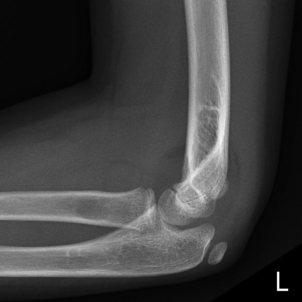
Case 12
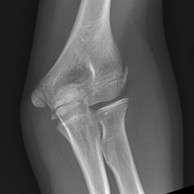
AP ELBOWThis 9 year old girl presented to the Emergency Department after falling off a chair. On examination, she was found to have a painful left elbow. She was referred for left elbow radiography.
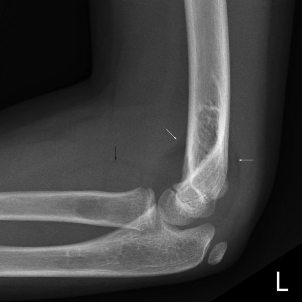
lateral elbowThe lateral elbow image demonstrates multiple soft tissue signs.
lateral elbowThere are anterior and posterior fatpad signs (white arrows).

There is a raised supinator fatpad (black arrow)
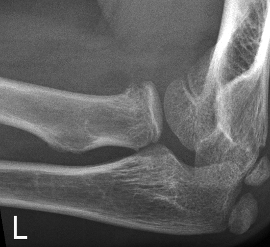
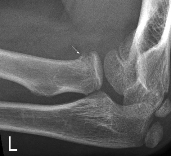
radial head projectionThis is a radial head projection image. There is irregularity of the proximal radial metaphysis.
radial head projectionThe arrowed irregularity of the proximal radial metaphysis probably represents a Salter Harris II fracture.


Summary

Patients with radial head fractures will occasionally provide a challenge for the trauma radiographer. Achieving good quality imaging in a patient who is in considerable pain can test the innovative/adaptive skills of junior and seasoned radiographers alike. Radial head fractures can be subtle requiring a balanced consideration of the mechanism of injury and subtle soft tissue signs when deciding which patients to pursue with supplementary views and which to save from further irradiation. Some radial head fractures will only be demonstrated radiographically on second or even third presentations.


... back to the Wikiradiography home page
...back to the Applied Radiography home page