---under construction---
IntroductionOlecranon fractures occur in the adult and paediatric population. This page considers imaging techniques and image interpretation related to olecranon fractures
Case 1This 50 year old lady presented to the Emergency Department after falling onto her right arm. Her right elbow was painful and swollen. She was referred for right elbow radiography.
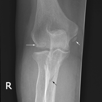 | This is an AP view image of the right elbow. There is a fracture of the right olecranon/ulna (black arrow). The corticated bony fragment adjacent to the medial condyle is likely to be an unfused ossification centre.
The nature of the medial condyle appearance (horizontal white arrow) is uncertain- a fracture cannot be excluded |
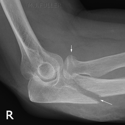 | This is a malpositioned lateral elbow image. The olecranon/ulna fracture is clearly demonstrated (large white arrow). In addition, there is evidence of a radial head/neck fracture (small white arrow) |
Comment
This fracture is typical of a fracture caused by a direct blow to the dorsal aspect of the forearm, usually from a fall. It is good radiographic practice to ensure that there are no other associated fractures- additional views should be performed if there is evidence of associated fractures which are not clearly demonstrated.
Case 2This 96 year old lady presented to the Emergency Department after falling directly onto her left elbow. She was in considerable pain and showed left elbow swelling. She was referred for radiography of her left elbow.
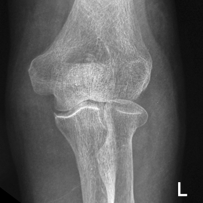 | The AP elbow view image suggests a possible olecranon fracture. |
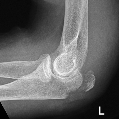 | The lateral view image confirms the olecranon fracture. |
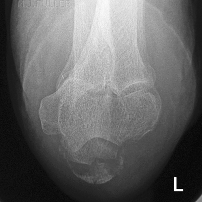 | The radiographer considered that an axial view of the elbow would provide additional information regarding the nature of the fracture and the degree of comminution. The axial view has been executed to good effect demonstrating the fracture effectively. |
Comment
This case demonstrates the potential value of the axial view of the elbow in cases of olecranon fracture.
Case 3This 51 year old man presented to the Emergency Department after falling onto both hands. He was examined and found to have a painful and deformed right elbow and was referred for radiography of his right elbow.
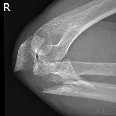 | The elbow is overflexed. It is likely that this is the position that the patient presented with and the radiographer correctly did not change the position for the initial series. There are fractures of the olecranon and radial neck. |
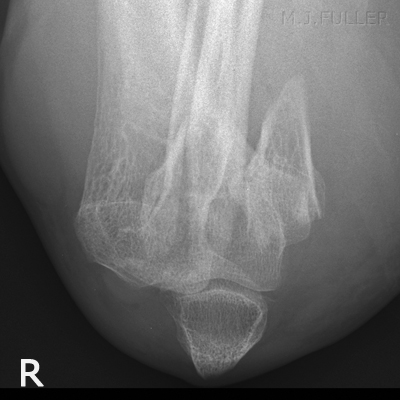 | Irrespective of whether the axial view was the ideal view for demonstrating these fractures, the patient wouldn't (and shouldn't) move his elbow and the radiographer correctly performed an axial view to provide the referring doctor with two views at 90 degrees with minimum patient discomfort. |
Case 4This 7 year old girl Presented to the Emergency Department after falling onto her left elbow . She was examined and found to have a painful and swollen left elbow and was referred for radiography of her left elbow.
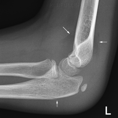 | The elbow is in a true lateral position.
The lateral elbow image demonstrates anterior and posterior fatpad signs indicating intra-articular injury. (top arrows)
A subtle cortical defect is noted in the dorsal aspect of the olecranon (bottom arrow) |
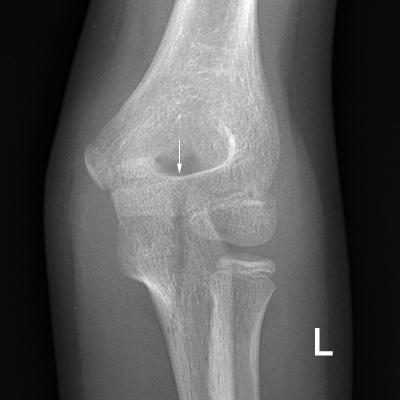 | The AP elbow view image demonstrates a fracture of the olecranon. (arrowed) |
Case 5This 38 year old man presented to the Emergency Department after an unknown injury . He was found to have a painful and swollen left elbow and was referred for radiography of his left elbow.
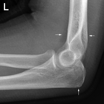 | The elbow is in an acceptable lateral position.
There are anterior and posterior fat pad signs indicating intra-articluar injury. (top white arrows)
There is a fracture of the olecranon process (bottom white arrow). |
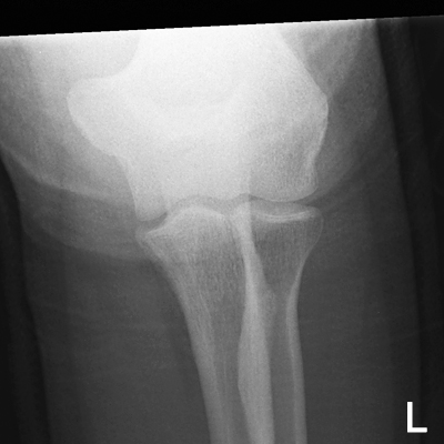 | The AP elbow view was performed with the forearm in contact with the IR. Whilst this is the better position for demonstrating the forearm anatomy, it has the disadvantage of potential underexposure of the upper arm (as in this case).
No displaced fracture is demonstrated. |
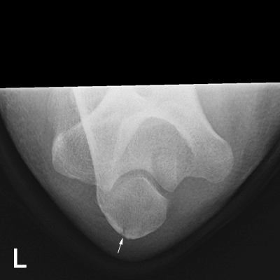 | An axial elbow view was performed. This demonstrated the distal olecranon that was not well demonstrated on the AP view because of underexposure. This view suggests that the fracture of the olecranon process is largely undisplaced. |
Case 6This 5 year old girl presented to the Emergency Department after falling from a railing onto her left arm. Her left elbow was very swollen and painful. She was referred for radiography of her left elbow.
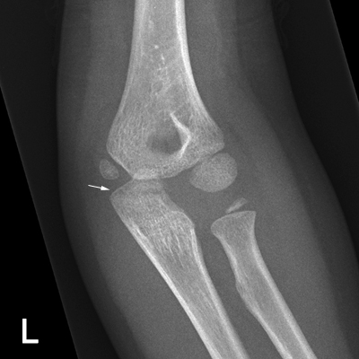 | There is a lucent line through the olecranon process indicating a fracture (white arrow). |
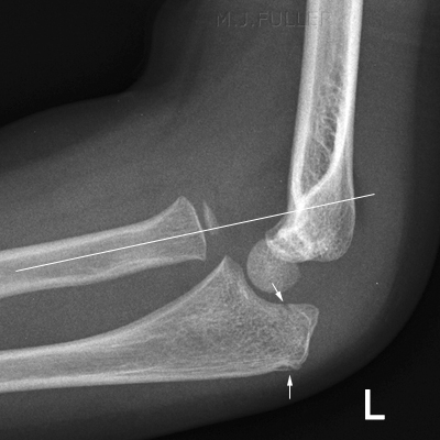 | There are anterior and posterior fatpad signs (not marked)
There is also further evidence of a fractured olecranon process (white arrows). The bottom white arrow is probably pointing to a normal structure given the lack of associated soft tissue swelling. The top arrow is more convincing of a fracture.
The lateral elbow image also demonstrates a radial head dislocation- the radial line does not bisect the capitellum. |
Comment
This case illustrates the need for care when assessing childen's elbow plain films. There can be more than one pathology. Beware satisfaction syndrome.
Case 7This 66 year old lady presented to the Emergency Department after falling onto her left elbow. Her left elbow was very swollen and painful and there was evidence of crepatis. She was referred for radiography of her left elbow.
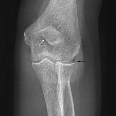 | This is an AP view image of the left elbow. There is an unusual appearance of the distal olecranon process (white arrow). There is also evidence of air in the soft tissues (subctaneous emphysema) and possibly in the elbow joint joint (black arrow). |
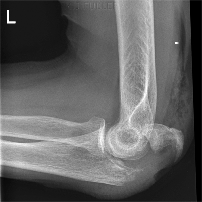 | The lateral view demonstrates a displaced olecranon fracture. There is also extensive subcutaneous emphysema (white arrow) |
Comment
Subcutaneous emphysema and air in the joint are important findings in MSK acute plain film radiography. Where there are subtle signs of air in the joint, additional views should be considered to confirm the finding.
...back to the
Wikiradiography home page
...back to the
Applied Radiography home page


