Elbow Medial Condyle Fractures
Mechanism of Injury
Separation (avulsion) of the medial epicondylar apophysis occurs in trauma or "... in child and adolescent athletes as a result of a single very strenuous throwing effort or as a result of repeated throwing motions that require valgus force at the elbow". <a class="external" href="http://books.google.com.au/books?id=oa6fDFuX-I8C&pg=PA523&lpg=PA523&dq=elbow+condyle+vs+epicondyle&source=bl&ots=lhiHITotg0&sig=YDhHTE7q_t_nQN0pcYGHEr0kFqo&hl=en&ei=wSBPSrS3Loj8sQPOvYWrDQ&sa=X&oi=book_result&ct=result&resnum=7" rel="nofollow" target="_blank">(Robert. Bruce. Salter ,</a><a class="external" href="http://books.google.com.au/books?id=oa6fDFuX-I8C&pg=PA523&lpg=PA523&dq=elbow+condyle+vs+epicondyle&source=bl&ots=lhiHITotg0&sig=YDhHTE7q_t_nQN0pcYGHEr0kFqo&hl=en&ei=wSBPSrS3Loj8sQPOvYWrDQ&sa=X&oi=book_result&ct=result&resnum=7" rel="nofollow" target="_blank"> Textbook. of Disorders and Injuries of the Musculoskeletal System. 3rd Ed, 1999, p344)</a>. The elbow has a normal valgus alignment. When a child falls on the outstretched arm, this can lead to an extreme valgus angular force. When the forces have more effect on the humerus, the extreme valgus will result in a fracture of the lateral condyle. On the medial side the valgus force can lead to avulsion of the medial epicondyle. Because of the valgus position of the normal elbow an avulsion of the lateral epicondyle will be uncommon. <a class="external" href="http://www.radiologyassistant.nl/en/4214416a75d87" rel="nofollow" target="_blank">(Robin Smithuis, The Radiology Assistant, Elbow - Fractures in Children)</a>
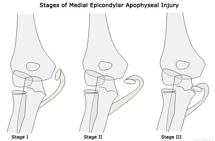
Stages of Medial Epicondyle Apophyseal Injury
based on Swischuk, Leonard E. Emergency Radiology of the Acutely Ill or Injured Child Williams & Wilkins; 2nd edition, January 1986The medial epicondyle is the third ossification centre to appear at the elbow and the last to fuse. <a class="external" href="http://www.imageinterpretation.co.uk/elbow.html" rel="nofollow" target="_blank">(Heidi Gable, Image Interpretation Course) </a>
Between the ages of 5 and 20 the centre of ossification of the medial epicondyle is a separate piece of bone. The flexor muscles of the forearm are attached to it, and if these are pulled on hard enough by a fall on an outstretched hand, they can pull it away from a patient’s humerus. His detached medial epicondyle may remain outside the elbow joint or rest inside the joint and lock it.
<a class="external" href="http://www.primary-surgery.org/ps/vol2/html/sect0385.html" rel="nofollow" target="_blank">http://www.primary-surgery.org/ps/vol2/html/sect0385.html</a>
"Unlike many fractures of the elbow, fractures of the medial epicondylar apophysis do not involve the joint surface or growth cartilage. The medial epicondyle is a posteromedial structure that serves as the origin of the flexor-pronator muscle mass as well as the medial collateral ligamentous complex. About 80% of medial epicondyle fractures occur in boys with a peak age in early adolescence. The mechanism of injury is typically an acute valgus stress to the elbow, although chronic injuries can occur in growing athletes" <a class="external" href="http://www.uphs.upenn.edu/ortho/oj/2002/html/oj15sp02p43.html" rel="nofollow" target="_blank">(Junichi Tamai et al, Pediatric Elbow Fractures: Pearls and Pitfalls, UPOJ, Volume 15 Spring 2002 Pages 43-51). </a>
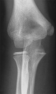
Normal
<a class="external" href="http://bp1.blogger.com/_nFuCC8zhFBc/SCbmkIT9bSI/AAAAAAAAArs/OeEmQYhDmX4/s1600-h/ScreenShot005.jpg" rel="nofollow" target="_blank">http://bp1.blogger.com/_nFuCC8zhFBc/SCbmkIT9bSI/AAAAAAAAArs/OeEmQYhDmX4/s1600-h/ScreenShot005.jpg</a>Stage I
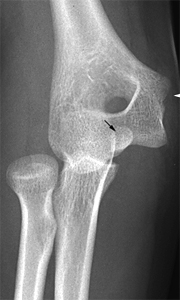
<a class="external" href="http://bp1.blogger.com/_nFuCC8zhFBc/SCbmkIT9bSI/AAAAAAAAArs/OeEmQYhDmX4/s1600-h/ScreenShot005.jpg" rel="nofollow" target="_blank">http://bp1.blogger.com/_nFuCC8zhFBc/SCbmkIT9bSI/AAAAAAAAArs/OeEmQYhDmX4/s1600-h/ScreenShot005.jpg</a>Stage II Stage III The medial epicondyle in a normal elbow should feature a smooth cortical contour from metaphysis to medial epicondyle. This is more commonly referred to as a Little Leaguer's Elbow. A significant diagnostic clue to this avulsion will be the associated medial soft tissue swelling. The danger with stage III avulsions is that the avulsed fragment can end up positioned within the elbow joint thus mimicking the trochlea. This is where the CRITOL rule becomes invaluable.
Case 1
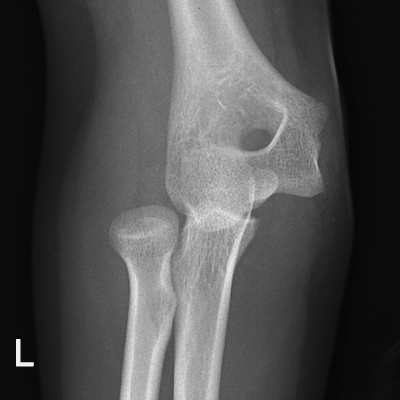
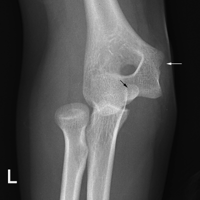
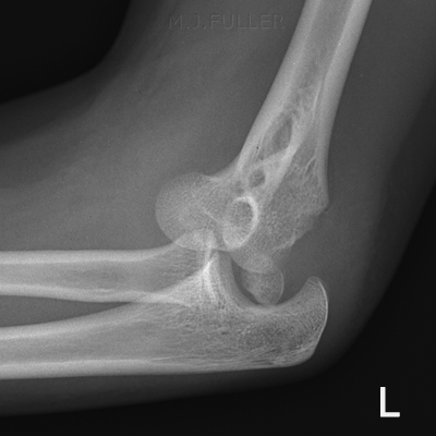
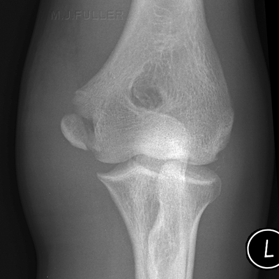
This 14 year old girl fell onto an outstretched left hand and reported that she thought her left elbow had "popped out". The left elbow was painful and deformed. She was referred for elbow radiography.
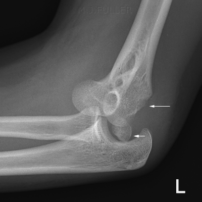
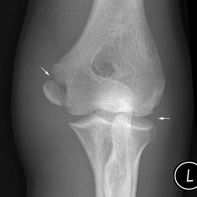
The radius and ulna are dislocated. The medial epicondyle has been avulsed (black arrow). The donor site is marked with a white arrow.
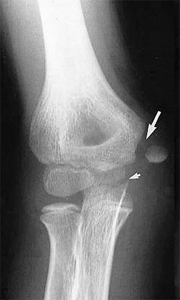
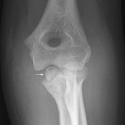
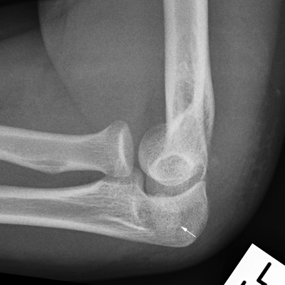
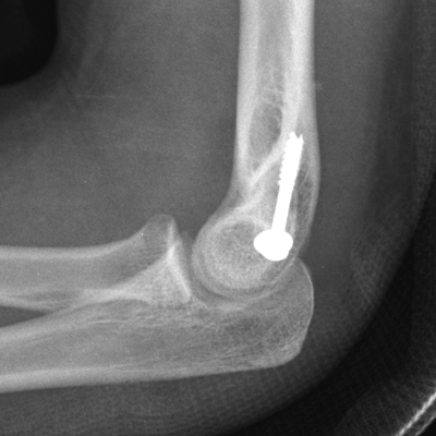
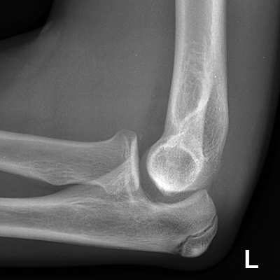
This is a stage III avulsion of the medial epicondyle in which the medial epicondyle has become sited within the elbow joint. This injury is known to be associated with elbow dislocation. There is approximately 50% incidence of associated elbow dislocations with medial epicondyle fractures <a class="external" href="http://www.uphs.upenn.edu/ortho/oj/2002/html/oj15sp02p43.html" rel="nofollow" target="_blank">(Junichi Tamai et al, Pediatric Elbow Fractures: Pearls and Pitfalls, UPOJ, Volume 15 Spring 2002 Pages 43-51).
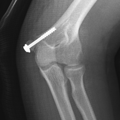
</a>The avulsed medial epicondyle (small white arrow) is clearly demonstrated along with the donor site (large white arrow) The post-reduction images demonstrate that the medial epicondyle remains located within the medial aspect of the elbow joint. Note also the increased distance between the ulna and the trochlea (compare with the post-fixation images below) Post fixation images demonstrate the medial epicondyle relocated and fixed with a screw.
Case 2
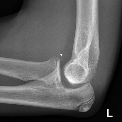
This 14 year old boy fell off a trampoline onto an outstretched left hand. The left elbow was painful and deformed. He was referred for elbow radiography. The original imaging demonstrated a posterior dislocation of the elbow. The patient was referred for post reduction radiography to ensure relocation and establish if there were any associated fractures.
Case 3
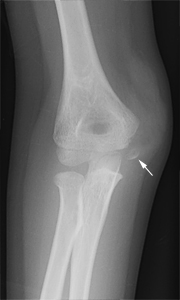
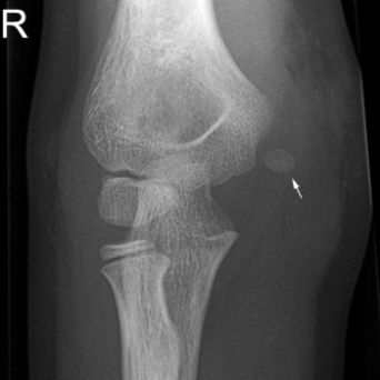
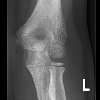
This 8 year old boy presented to the Emergency Department with a painful swollen right elbow. The mechanism of injury was unknown.
...back to the Wikiradiography home page
...back to the Applied Radiography home page
