Axial Elbow Radiography

From wikiRadiography
Jump to navigation Jump to search
Introduction

The axial view of the elbow is one of those views that stands as a hallmark of innovative radiographers. The radiographer who utilizes this view when required is usually accomplished in plain film radiographic technique.

Radiography
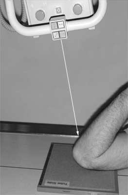
axial elbow position
adapted from <a class="external" href="http://ajs.sagepub.com/content/31/3/466/F1.large.jpg" rel="nofollow" target="_blank">http://ajs.sagepub.com/content/31/3/466/F1.large.jpg</a>
The essence of the technique is that you can achieve a satisfactory demonstration of the distal humerus in an AP projection by superimposing the forearm while the elbow is flexed.


Case 1
axial elbowaxial elbow
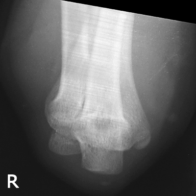
This is an image from circa 1958. The age of the patient and the mechanism is unknown. The olecranon epiphysis is faintly visible which would provide an estimate of the child's age at around 11 years. This is a (pointless) comparison axial view.

Note the wash marks associated with manual processing.
The left axial elbow image demonstrates a fracture of the lateral condyle.


Case 2

This 96 year old lady presented to the Emergency Department after falling directly onto her left elbow. She was in considerable pain and showed left elbow swelling. She was referred for radiography of her left elbow.

AP elbowThe AP elbow view image suggests a possible olecranon fracture.
lateral elbowThe lateral view image confirms the olecranon fracture.
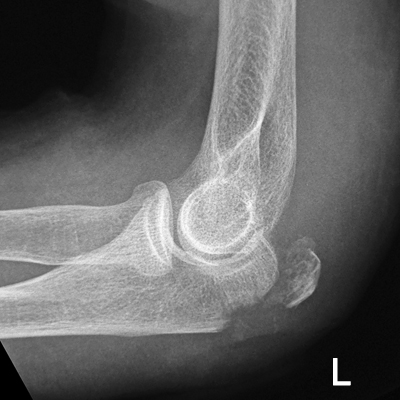
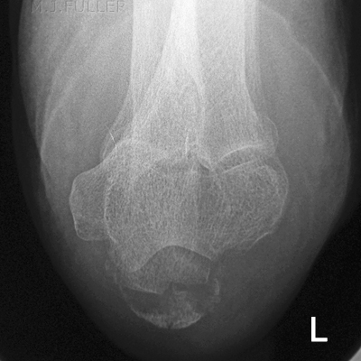
axial elbowThe radiographer thought that an axial view of the elbow would provide additional information regarding the nature of the fracture and the degree of comminution. The axial view has been executed to good effect demonstrating the fracture effectively.


Case 3

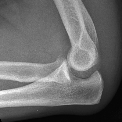
This 7 year old girl presented to the Emergency Department after an unwitnessed fall. She was referred for radiography of her left elbow.

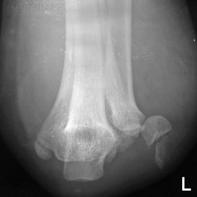
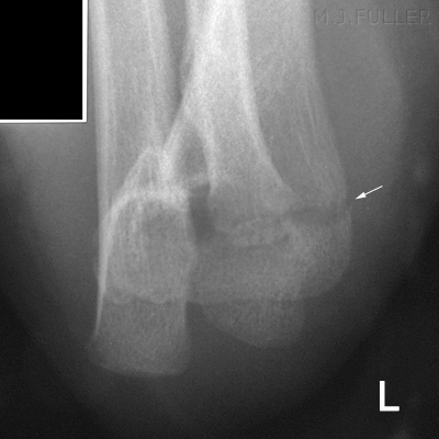
axial elbowThe axial elbow view was performed in lieu of the conventional AP view.
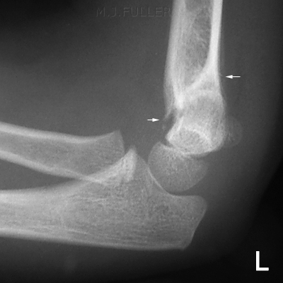
lateral elbowThe lateral view image confirms the supracondylar fracture.


Case 4

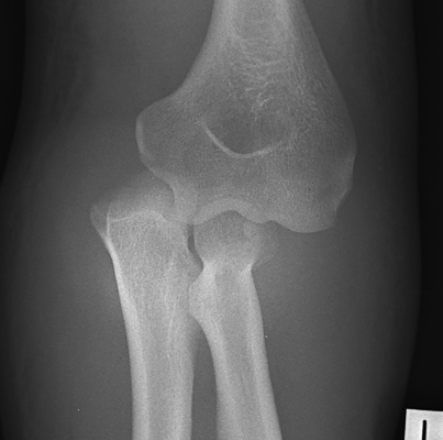
This 34 year old man presented to the Emergency Department after falling from a roof. He was examined and found to have a painful and deformed left elbow and was referred for radiography of his left elbow.

lateral elbowThe lateral elbow view is underexposed and malpositioned. There was no obvious abnormality
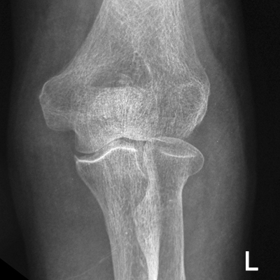
axial elbowComparison View
axial elbow
Compare with the normal axial elbow above.
The axial view shows the olecranon to be dislocated.
AP elbowThe AP elbow view demonstrates that the olecranon and radial head are dislocated. There is also evidence of a radial head fracture


Case 5

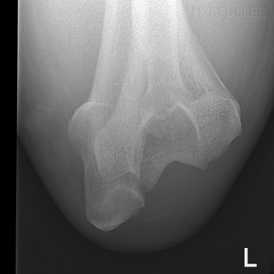
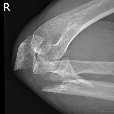
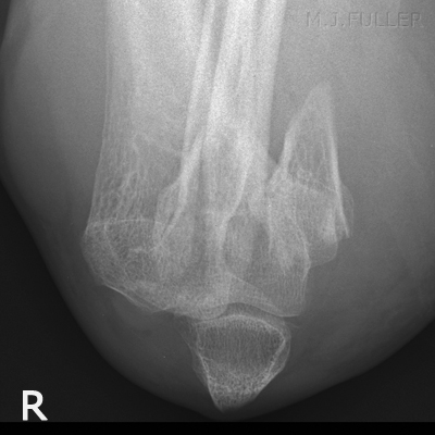
This 51 year old man presented to the Emergency Department after falling onto both hands. He was examined and found to have a painful and deformed right elbow and was referred for radiography of his right elbow.

lateral elbowThe elbow is overflexed. It is likely that this is the position that the patient presented with and the radiographer correctly did not change the position for the initial series. There are fractures of the olecranon and radial neck producing an unstable elbow joint.
axial elbowIrrespective of whether the axial view was the best view for demonstrating these fractures, the patient wouldn't (and shouldn't) move his elbow and the radiographer correctly performed an axial view to provide the referring doctor with two views at 90 degrees.


Case 6

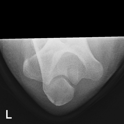
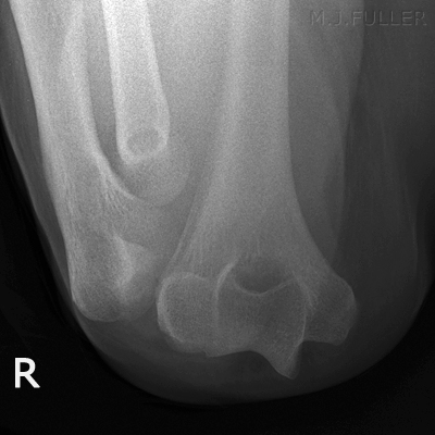
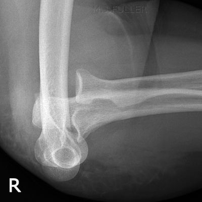
This 70 year old lady presented to the Emergency Department after falling onto her right side. She was examined and found to have a painful and deformed right elbow and was referred for radiography of her right elbow.

AXIAL ELBOWThe patient was in considerable pain and distress with a fixed flexed elbow. The radiographer chose to perform an axial view as a first view in the series. The axial view image demonstrates complete dislocation of the elbow joint.
LATERAL ELBOWThe lateral view similarly shows complete disruption of the elbow joint with 90 degree rotation of the radius and ulna with respect to the humerus.


...back to the Wikiradiography home page
...back to the Applied Radiography home page