The Paediatric Elbow

From wikiRadiography
Jump to navigation Jump to search

Introduction

In many respects paediatric elbow radiography can be a challenge. A perfect lateral elbow projection can be difficult to achieve and this difficulty is exacerbated in a child who is scared and in pain. This page looks at what you are trying to achieve and what to look out for.


The Lateral Elbow

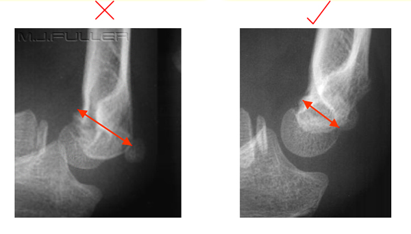
The lateral elbow projection, paediatric or adult, is one of the most difficult views in radiography. If you have achieved the correct position, the elbow will be flexed to 90 degrees, the patient's wrist will be in the lateral position, and the distal humerus will have a minimum antero-posterior dimension. The distal humerus is wider in its lateral dimension than in its AP dimension. Therefore, if you start with a perfectly lateral elbow, and you rotate it off lateral, the distal humerus it will increase in its AP dimension. In other words, a true lateral elbow will demonstrate the distal humerus at its narrowest.


Minimal Antero-posterior (AP) Distance
The Paediatric Elbow - wikiRadiographyThe true lateral elbow has a minimal AP dimension.

The medial epicondylar apophysis is projected almost clear of the distal humerus on the malpositioned view. This suggests that the patient's hand needs to be raised to correct this malposition.


The Hockey Stick Analogy

The Paediatric Elbow - wikiRadiography

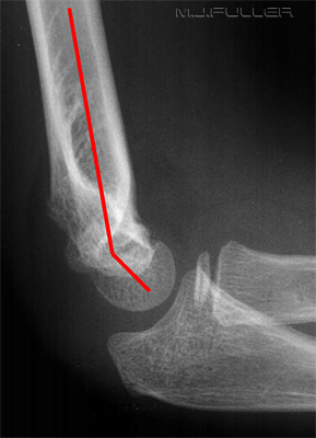
The lateral elbow has been likened to a hockey stick shape as shown left.

Whilst it is undeniable that this lateral elbow looks hockey stick-shaped, this is not a useful aid in assessing lateralness. A lateral elbow can be very "off-lateral" and still retain the hockey stick shape.


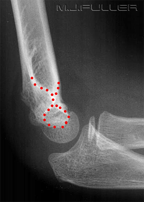
The Figure Eight or Hour Glass Sign
First Attempt
The Paediatric Elbow - wikiRadiography
Second Attempt
The Paediatric Elbow - wikiRadiography
Second Attempt
The Paediatric Elbow - wikiRadiography
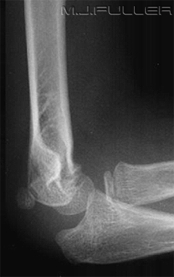
This is the first attempt at a lateral elbow.
Note the absence of the figure eight sign. Note also the medial epicondylar apophysis projected posteriorly.
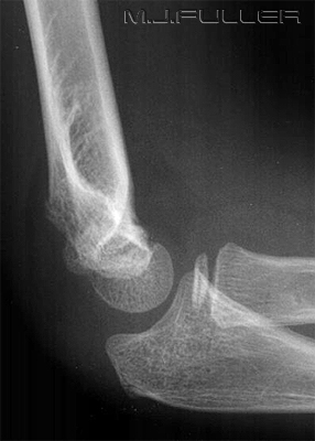
This is the second attempt at the lateral elbow.
The patient's wrist has been raised slightly. This movement has caused the medial epicondylar apophysis to be partially superimposed over the distal humerus and the radial head to further overlap the ulna.
This is logical if you think about the relationship between the anatomical structures.

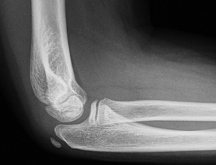
A better position has been achieved and the
figure
eight sign is now evident

Note a subtle supraconylar fracture
Second attempt (same image) with figure eight sign marked


Important

The figure eight sign does not indicate a perfectly positioned lateral elbow. However, the absence of the figure eight sign could indicate that the elbow is not lateral, or that the elbow is fractured, or both.


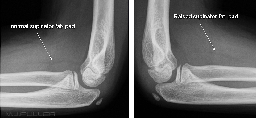
The Supinator Fatpad Sign
The Paediatric Elbow - wikiRadiography
The supinator fat pad can be raised or obliterated as a result of bony injury, particularly to the radial neck.
It is one of those unreliable soft tissue signs, but it is worth examining as a guide to potential bony injury, particularly to the radial neck.


Tip: Supinator or Pronator Memory Aid?

<a class="external" href="http://globalvillagebrisbane.files.wordpress.com/2008/12/waiter_-_cartoon_4.jpg" rel="nofollow" target="_blank">supinator cartoon</a>

<a class="external" href="http://globalvillagebrisbane.files.wordpress.com/2008/12/waiter_-_cartoon_4.jpg" rel="nofollow" target="_blank">Adapted from http://globalvillagebrisbane.files.wordpress.com/2008/12/waiter_-_cartoon_4.jpg</a>

If you have trouble remembering whether it is the supinator fat pad sign or the pronator fat pad sign, think of a restaurant waiter. Waiters sometime carry soup bowls (i.e. "soupinator") in their elbows.
..... pronator fat pad at the wrist.... supinator fat pad at the elbow.


The Fat Pad Sign

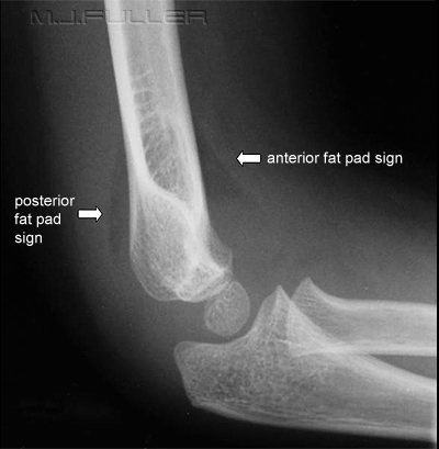
The Paediatric Elbow - wikiRadiographyThe fat pad sign or sail sign indicates that the patient has sustained an intra-articular injury. Importantly, it does not indicate that the patient has definitely sustained an intra-articular bony injury.


i.e. fat pad sign does not equal fracture

fat pad sign does indicate in increased chance of a fracture


There have been studies investigating the correlation between the fat pad sign and bony injury. These studies have reported an anterior fat pad sign indicates a probability of intra-articular fracture of approximately 70-80%, whereas a posterior fat pad sign indicates a 90% chance of intra-articular fracture. The anterior fat pad can be seen in a normal elbow. The posterior fat pad sign is never visible in a normal elbow.


Anatomy
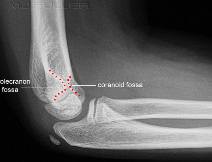
The Paediatric Elbow - wikiRadiographyThe Paediatric Elbow - wikiRadiography


In a normal elbow, anterior fat pad is nestled in the coronoid fossa. The posterior fat pad occupies the olecranon fossa.The olecranon fossa is larger to accommodate the olecranon, and the coronoid fossa is smaller to accommodate the coronoid process. This is consistent with the observation that a larger elbow joint effusion is required to reveal the posterior fat pad. Note that this patient has a visible normal anterior fat pad.

Don't get the coracoid of the shoulder confused with the coronoid of the elbow.


The Normal Elbow Alignment lines
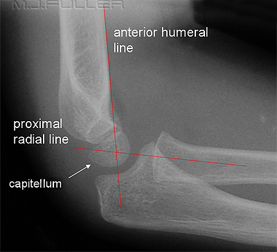
lateral elbowThe normal alignment lines of the elbow can be a guide both to good positioning and the existence of subtle pathology. The two normal alignment criteria are the anterior humeral line and the proximal radial line as shown left.

The anterior humeral line is a line drawn along the anterior cortex of the distal humerus in the lateral view. This line is described as passing through the middle third of the capitellum. If it is clearly not in this position, it suggests that the elbow is not lateral or that there is a fracture.


The proximal radial line is a line drawn through the middle of the radius that should bisect the capitellum. If it does not divide the capitellum into halves, it could indicate a dislocated radial head. Don't get too picky as to whether it exactly divides the capitellum in half- it depends how you draw the line.

Another useful feature of the proximal radial line is that it should always bisect the capitellum in any projection.


Abnormal Proximal Radial Line Elbow Alignment
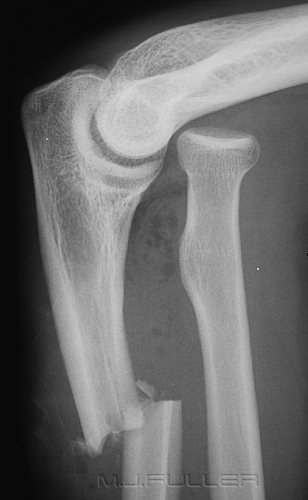
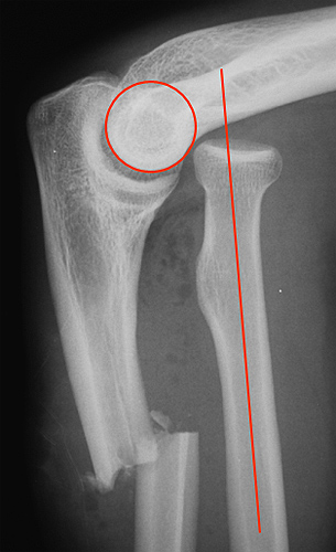
The Paediatric Elbow - wikiRadiographyThe Paediatric Elbow - wikiRadiography
The proximal radial line does not line up with the middle of the capitellumNote that you cannot see the capitellum, but you know its
approximate position and that the proximal radial line is
not even close


Abnormal Anterior Humeral Line Elbow Alignment
The Paediatric Elbow - wikiRadiography
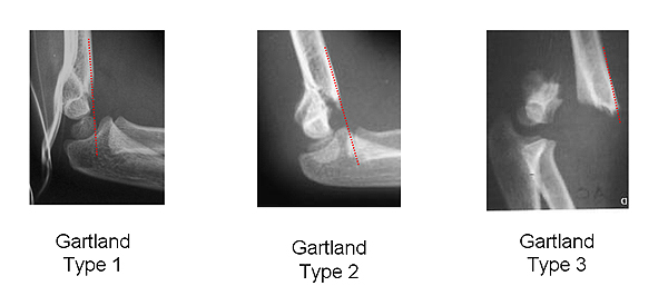
Note that the anterior humeral line does not divide the capitellum into thirds in any of these lateral elbow images. See further down this page for more information about the Gartland classification of supracondylar fractures.


The CRITOL Rule

ossification of the elbow

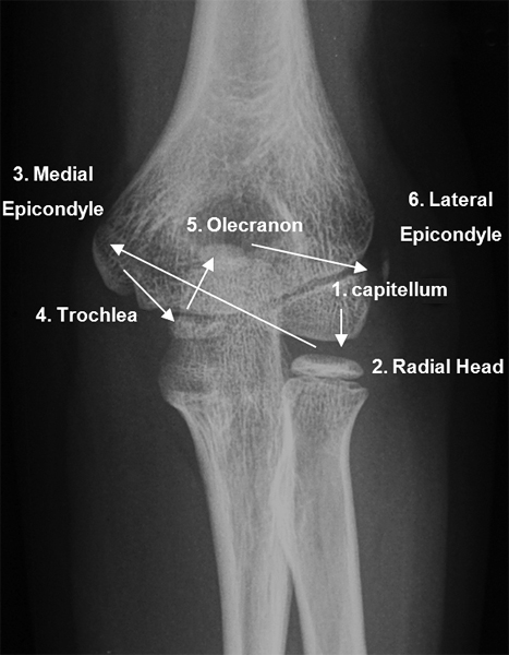
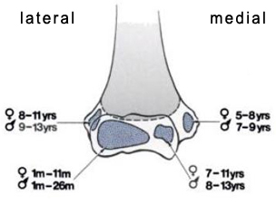
The appearances of the ossification centres of the elbow frequently causes confusion. The CRITOL rule is a memory aid that lists the order of appearance of the elbow ossification centres


The order of appearance of the elbow ossification centres is as follows

1. Capitellum
2. Radial Head
3. Internal (medial epicondyle)
4. Trochlea
5. Olecranon
6. Lateral Epicondyle


The capitellum contributes to the growth of the humerus and is therefore considered an epiphysis. The other ossifications centres are called traction epiphyses or apophysis.

Some Important Notes about the CRITOL Rule
  • It is not uncommon for the ossification centres to appear out of order
  • The ages at which the ossification centres appear is approximate only. Different texts will suggests different ages
  • The trochlea often appears fragmented- this is normal
  • the "I" in CRITOL refers to the medial epicondylar ossification centre


Case 1
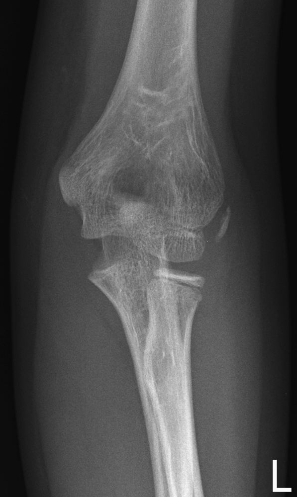
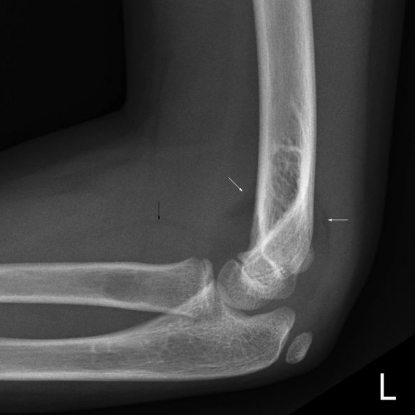
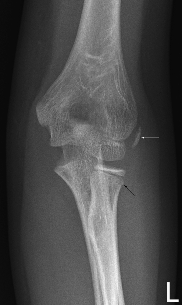
AP ELBOWThis 9 year old girl presented to the Emergency Department after falling off a chair. On examination, she was found to have a painful left elbow. She was referred for left elbow radiography.
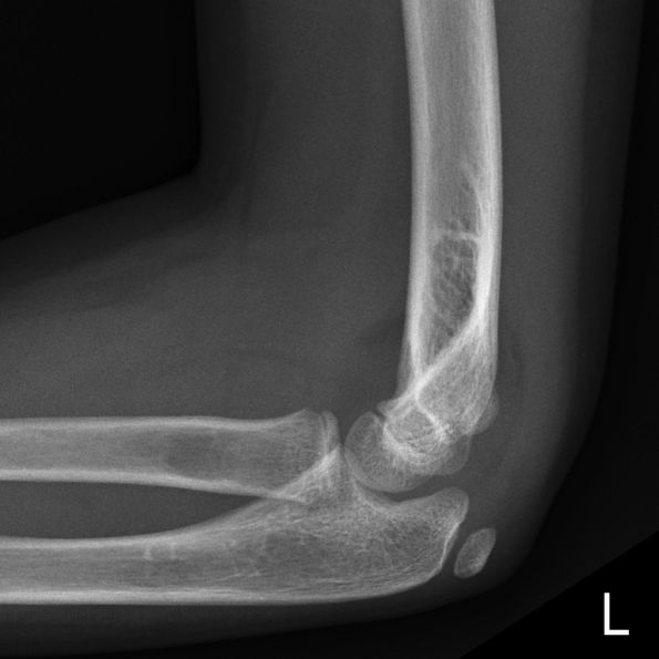
lateral elbowThe lateral elbow image demonstrates multiple soft tissue signs.
lateral elbowThere are anterior and posterior fatpad signs (white arrows).

There is a raised supinator fatpad (black arrow)
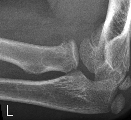
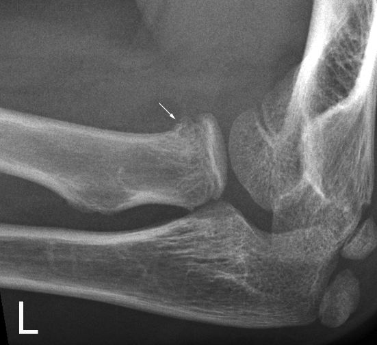
radial head projectionThis is a radial head projection image. There is irregularity of the proximal radial metaphysis.
radial head projectionThe arrowed irregularity of the proximal radial metaphysis probably represents a Salter Harris II fracture.


Discussion

AP elbowThe SHII fracture (black arrow) was demonstrated on the AP elbow projection image (albeit subtle).

Is the white arrowed structure
  • a normal lateral apophysis?
  • an avulsed lateral apophysis?
  • an lateral epicondylar avulsion fracture?


The radiographer considered it to be a normal structure; the referring doctor considered it to be a normal structure; the orthopaedic surgeon considered it to be an avulsion fracture; the radiology registrar considered it to be normal and so did the consultant radiologist.. they cant all be correct!

CRITOL Rule

elbow ossification
<a class="external" href="http://books.google.com.au/books?id=6AXkE4y1678C&pg=PA509&lpg=PA509&dq=radial+head+fractures+incidence+in+adults&source=bl&ots=JRFuoZ23Kh&sig=r6wMfpmEAfwn6_ySqvaTdogcJVk&hl=en&ei=u_FHSvmRIIfU7AOqkZnoAw&sa=X&oi=book_result&ct=result&resnum=9" rel="nofollow" target="_blank">Charles A. Rockwood, Kaye E. Wilkins, James H. Beaty, James R. Kasser
Rockwood and Wilkins' fractures in children
Lippincott Williams & Wilkins, 2006</a>

The application of the CRITOL rule suggests an avusion fracture. The lateral ossification centre should follow the ossification of the trochlea. The trochlea is not yet convincingly ossified (although may show early signs). The elbow ossification centres are known to ossify out of order.The lateral ossification centre should appear in the 8-11 agegroup. This child is 9 years old.


Mechanism of Injury

elbow fracture mechanism

elbow fracture mechanism of injury<a class="external" href="http://www.radiologyassistant.nl/en/4214416a75d87" rel="nofollow" target="_blank">http://www.radiologyassistant.nl/en/4214416a75d87</a>

The mechanism of injury was a fall. It is possible that the child attempted to break her fall with an outstretched hand. This mechanism would explain the proximal radius fracture. This mechanism is consistent with an avulsion of the medial epicondyle not the lateral epicondyle.

Soft Tissue Signs

The soft tissues adjacent to the lateral epicondyle appear normal. Elbow avulsion fractures in children should demonstrate associated changes in the adjacent soft tissues.


... back to the Applied Radiography home page