Lateral Chest Case 6
Jump to navigation
Jump to search
Preamble
Other Relevant Wikiradiography Pages
Comment
... back to the wikiRadiography home page
... back to the Applied Radiography home page
... back to the What is the Value of the Lateral Chest Projection? page
This is the answer page to Case 6 from the page titled What is the Value of the Lateral Chest Projection?
Other Relevant Wikiradiography Pages
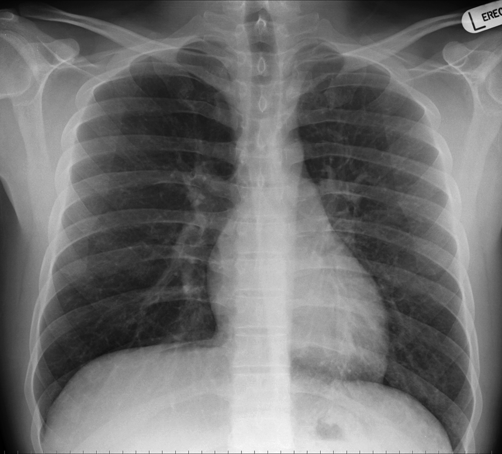
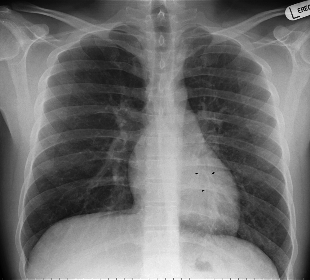
PA Erect Projection
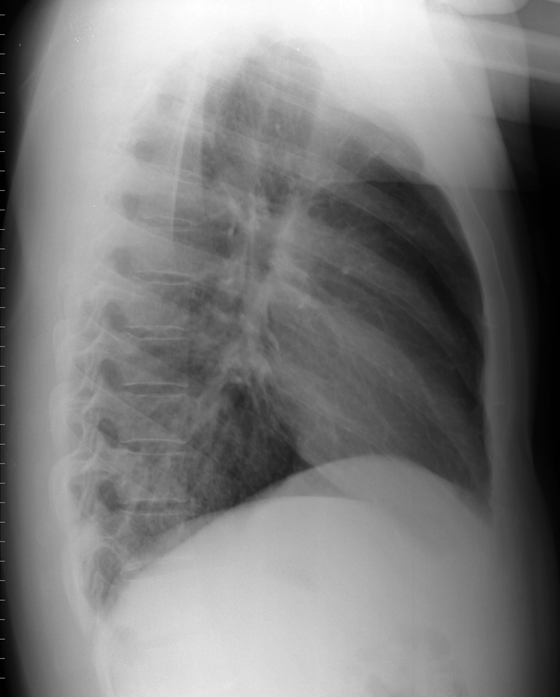
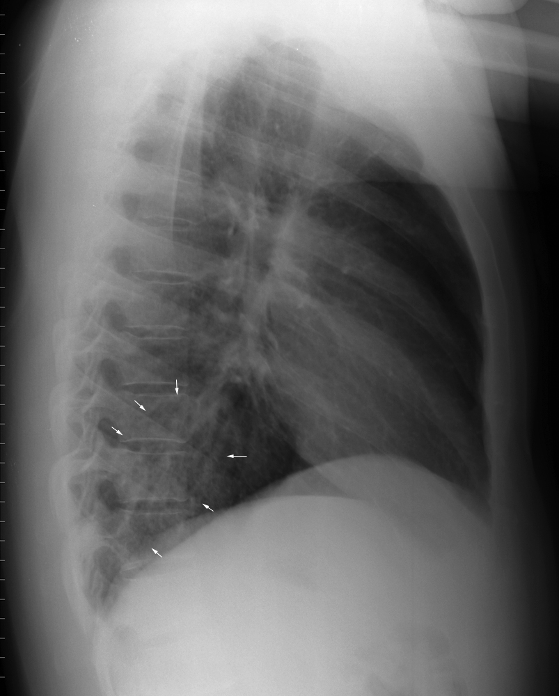
Lateral Projection
There is prominence of the posterior basal retrocardiac lung markings
Comment
This patient's PA chest findings were subtle (although they were noted by the radiographer). The lateral chest projection image provided additional confidence to the diagnosis and may have been influential in treatment decisions. It is at least possible (if not likely) that this finding would have been missed in an after-hours situation where no radiologist was present. I would also suggest that the risk of misdiagnosis would be higher if a lateral projection image was not provided.
... back to the wikiRadiography home page
... back to the Applied Radiography home page
... back to the What is the Value of the Lateral Chest Projection? page


