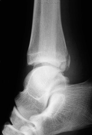
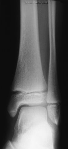
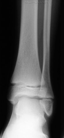
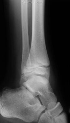
Lateral Ankle Radiography
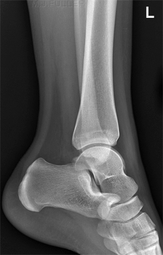
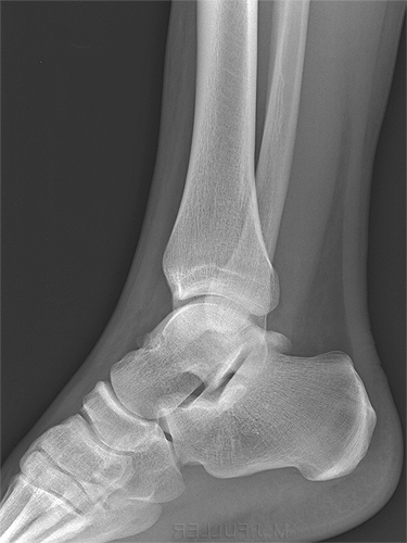
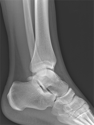
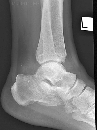
The lateral ankle position is not difficult to achieve radiographically. An understanding of normal anatomy, normal anatomical variants, soft tissue signs, positioning techniques and positioning errors can assist in achieving the best possible diagnostic results.
Indications for Ankle Radiography
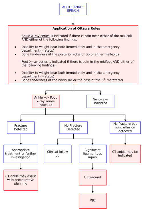
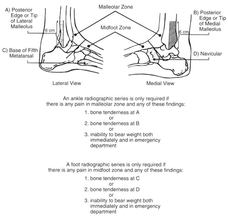
Patients with ankle soft tissue injuries are often inappropriately referred for ankle radiography . The most effective method of avoiding referrals for inappropriate ankle radiography is to have your institution sanction an evidence-based ankle imaging pathway. These decision pathways are commonly based on the Ottawa Rules. The following referral guidelines for ankle imaging are taken from an excellent series produced by the Western Australian Department of Health.
Source: <a class="external" href="http://www.imagingpathways.health.wa.gov.au/includes/pdf/ankle.pdf" rel="nofollow" target="_blank">Western Australian Department of Health</a>
Ottawa ankle rules for foot and ankle radiographic series in acute ankle injury patientsadapted from
Stiell IG, Greenberg GH, McKnight RD, et al.
Decision rules for the use of radiography in acute
ankle injuries: refinement and prospective validation.
JAMA. 1993;269:1127-1132.
in <a class="external" href="http://jama.ama-assn.org/cgi/reprint/271/11/827" rel="nofollow" target="_blank">http://jama.ama-assn.org/cgi/reprint/271/11/827</a>
Positioning Tips for Lateral Ankle Radiography
- Slightly flex the patient's knee
- Dorsiflex the patient's ankle
- Employ a horizontal ray technique if required
- Include the head of fibula in the series if there is a possibility of Maisonneuve fracture
- Include the base of 5th metatarsal, particularly in cases of ankle inversion injury
- repeat the lateral ankle view if malpositioned
- Centre the X-ray beam to the ankle Joint
- Lower leg radiography is not ankle radiography- a tib/fib image will not pass for dedicated ankle radiography
How Lateral is Lateral?
adapted from
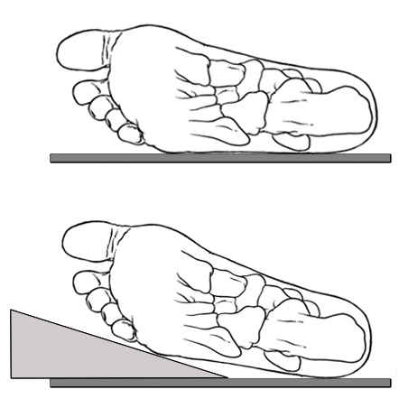
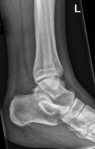
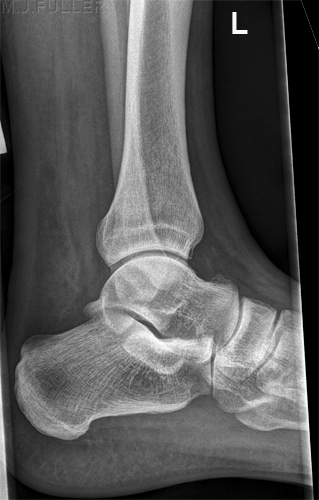
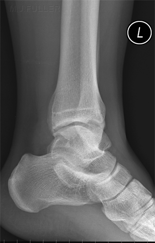
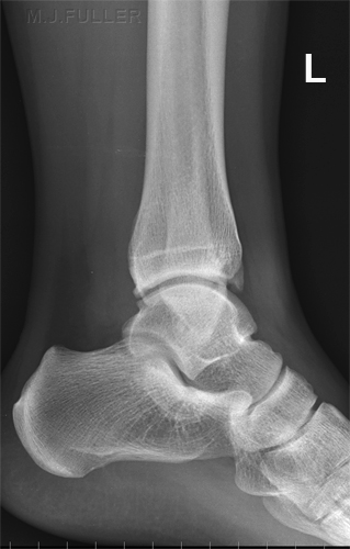
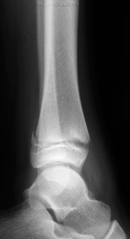
<a class="external" href="http://books.google.com.au/books?id=NePk5A1Y1NAC&printsec=frontcover" rel="nofollow" target="_blank">Skeletal Radiography: A Concise Introduction to Projection Radiography</a><a class="external" href="http://books.google.com.au/books?id=NePk5A1Y1NAC&printsec=frontcover" rel="nofollow" target="_blank">By Sheila Bull</a>The lateral ankle radiography challenge is largely a matter of deciding how far to roll the patient's ankle or, more specifically, how much to raise the patient's toes (if at all). This is not an easy judgement- consideration should be given to the width of the patient's foot. If you correctly position the lateral ankle, the medial and lateral talar domes will be overlapping and the joint will appear in profile. The exact position of the fibula is relation to the tibia in the true lateral ankle position will vary between patients, however, the position of the fibula slightly posteriorly in relation to the tibia is characteristic of a well positioned lateral ankle.
Correcting a Malpositioned Lateral ankle?
Case 1
Case 2
Soft Tissue Signs
Ankle Effusion
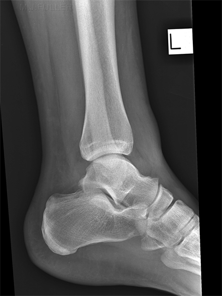
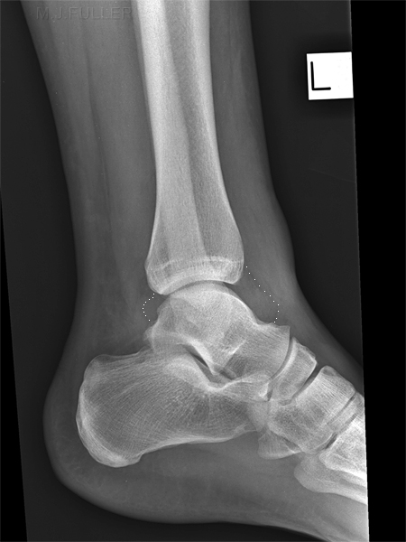
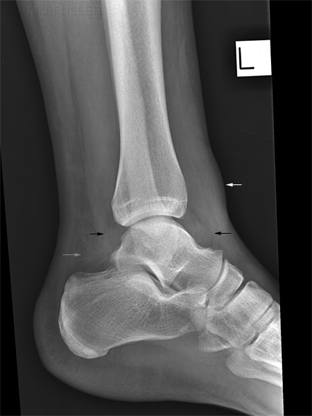
This lateral ankle image demonstrates
- ankle effusion (black arrows)
- extraarticular swelling anteriorly (white arrow)
- abnormal opacification of Kager's fatpad (grey arrow)
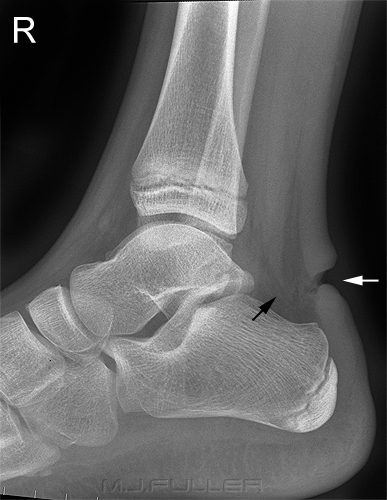
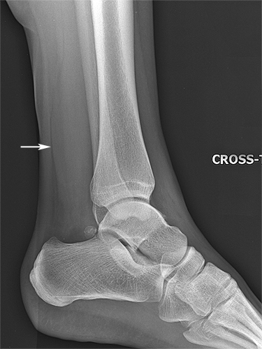
This patient has a ruptured Achilles tendon (white arrow). Note the changes in Kager's Fat Pad (black arrow) Normal Kager's fat pad with clearly delineated normal Achilles tendon Complete disruption of the Achilles tendon is most commonly seen in athletes and in men around the age of forty. Achilles tendon rupture can be treated surgically, or by placing the patient in a cast with equinus (marked plantar flexion) for several months. (1)
"Achilles tendon injuries- Physicians miss injuries to the Achilles tendon in 25% of cases, most often due to preservation of foot plantar flexion by the posterior tibial, peroneal, and toe flexor muscles? Patients will often describe the sensation of being kicked or “shot” in the back of the ankle with an immediately ensuing intense pain. Physicians should look for a palpable gap in the tendon and a positive Thompson test, since these are readily apparent in patients with complete rupture. With the patient in a prone position, instruct the patient to flex his knee. When the calf muscles are squeezed against the tibia and fibula, mechanical contraction of the gastrocnemius and soleus muscles occurs. If the Achilles tendon is ruptured, then contraction of the calf muscles will not plantar flex the foot. Incomplete ruptures may present greater diagnostic difficulties. In patients with these injuries, posteriorly located pain, swelling, and ecchymosis may be the only clue."
qouted from <a class="external" href="http://emcrit.org/030-064/051-ankle.foot.htm" rel="nofollow" target="_blank">http://emcrit.org/030-064/051-ankle.foot.htm</a>
References
(1) Helms, Clyde A. Fundamentals of Skeletal Radiology.3rd ed. Elsevier Saunders 2005
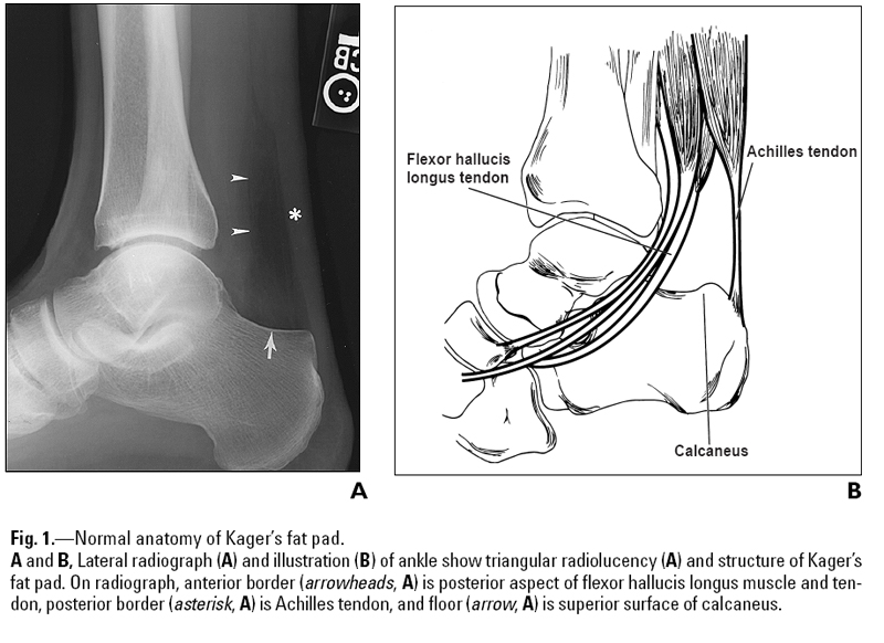
It is not uncommon for ankle injuries to involve Kager's fat pad. A careful examination of the density, shape and borders of Kager's fat pad can provide indicators of bony injury to the ankle. An abnormal Kager's fat pad does not indicate definite bony injury to the ankle.
<a class="external" href="http://www.ajronline.org/cgi/content/full/182/1/147" rel="nofollow" target="_blank">Justin Q. Ly and Liem T. Bui-Mansfield Anatomy of and Abnormalities Associated with
Kager’s Fat Pad AJR:182, January 2004</a>
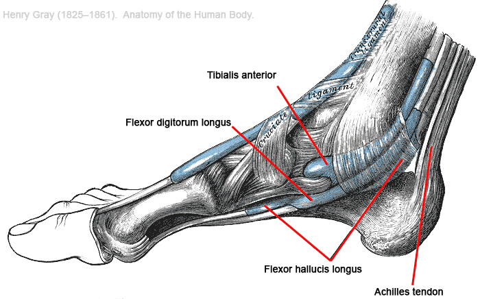
Further detail of ankle anatomy shown below
Case 1
Case 2
DiscussionIt is worth considering the risks of obscuring a posterior malleolus fracture of the distal tibia in cases where the lateral ankle position is incorrect as shown in the two cases above. Where there is a high suspicion index of a fracture based on patient history, clinical presentation and soft tissue signs ( e.g. ankle effusion) a repeat view of the lateral ankle is highly recommended.
Boehler’s Angle (aka Bohler's Angle)
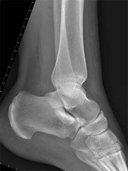
Lateral ankle radiography will usually include the calcaneum. Some patients who are referred for ankle radiography will have sustained a calcaneal fracture.
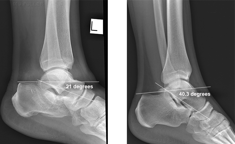
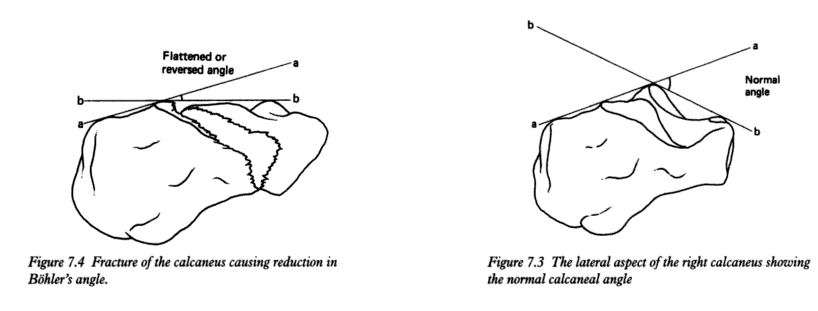
"Subtle [calcaneal] fractures may only be identified by assessing Boehler’s angle. This angle is measured by drawing a line from the highest point of the posterior tuberosity to the highest midpoint, and a 2nd line from the highest midpoint to the highest point of the anterior process. The angle, posteriorly, should be >30 degrees. If there is flattening of the bone due to a fracture, this angle will be decreased, to <30 degrees." <a class="external" href="http://imageinterpretation.co.uk/ankle.html" rel="nofollow" target="_blank">http://imageinterpretation.co.uk/ankle.html</a>
adapted from<a class="external" href="http://books.google.com.au/books?id=NePk5A1Y1NAC&printsec=frontcover" rel="nofollow" target="_blank">Skeletal Radiography: A Concise Introduction to Projection Radiography</a><a class="external" href="http://books.google.com.au/books?id=NePk5A1Y1NAC&printsec=frontcover" rel="nofollow" target="_blank">By Sheila Bull</a>The first image demonstrates a Boehler's Angle of 21 degrees suggesting that there is a fracture of the calcaneum. Compare this with the normal anatomy on the right. Note also the abnormal Kager's fatpad.
Normal Anatomical Variants
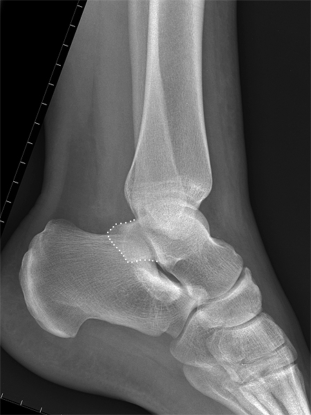
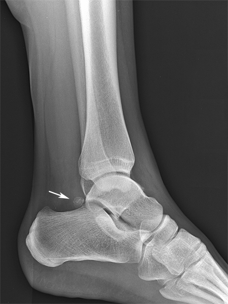
The most commonly seen normal anatomical variant on lateral ankle images is the os trigonum. This should not be confused with a fracture of the posterior process of the talus. Fractures of the posterior process of the talus are rare- the posterior process of the talus is somewhat protected by the Achille's tendon.
This patient has an unusually large posterior process of the talus Large posterior process of the talus marked
Os trigonum is variable in its size and shape (arrowed)
....back to the applied radiography home page here
