Soft Tissue Signs in Orthopaedics
Introduction
Health professionals with expertise in plain film image interpretation will approach an equivocal fracture by critically examining the bony features in the context of other information such as
- Patient history
- Clinical presentation
- soft tissue signs
This page will examine the soft tissue signs of bony injury- what they are, their plain film appearances, their limitations, and their utility.
Soft Tissue Signs.
Curtis D.J. and Downey E.F. (Gilula, L.A. The Traumatised Hand and Wrist, Radiographic and Anatomic Correlation, 1992, p50) observed the following in relation to radiography of the wrist.
| Soft tissue swelling involving fat planes should conform to the area of underlying soft tissue or bone injury. "Swelling seen in an area not associated with an observed fracture should initiate a search for an additional abnormality to explain the swelling. The possibility of a fracture or dislocation must be excluded with particular care, even though the routine views of the bones are normal in the presence of soft tissue swelling if more than one fat plane is affected or if the soft tissue swelling is unequivocal. The absence of soft tissue swelling virtually excludes the presence of a fracture, but the converse is not necessarily true... With definite swelling, additional views to better demonstrate the area underlying the swelling should be considered" (Gilula, L.A. The Traumatised Hand and Wrist, Radiographic and Anatomic Correlation,, 1992, p50). | ||
The Knee
Knee Effusion- Very Small
This patient presented with a history of a fall the previous day. There appears to be a small amount of fluid in the supra-patella pouch (left arrow). There is also fluid in Hoffa's Triangle (right arrow). Note that the knee is over-flexed. This position makes assessment of fluid in the supra-patella pouch more difficult.

Knee Effusion- Small
This patient presented with a history of dislocated right patella. There is a small knee effusion. There is a small amount of fluid in the supra-patella pouch (left arrow) and in Hoffa's Triangle (right arrow).

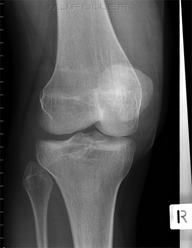
Knee Effusion- Moderately Large
The patient fell out of a tree onto both feet. There is a knee joint effusion. This is evident from the fluid density seen in the supra-patella pouch (left arrow)and in Hoffa's fat pad (right arrow). A knee effusion indicates an intra-capsular injury (but not necessarily a fracture). A knee effusion is not particularly specific or diagnostic, but the absence of a knee effusion can be useful in cases of equivocal fracture.

Knee Effusion- Very Large
This elderly patient fell onto her right knee. She has sustained a fractured patella and has a very large knee joint effusion (arrowed).
Extra-capsular Swelling
This patient's history is unknown. There is evidence of soft tissue swelling on the medial aspect of the knee on the AP image (arrowed). There is also soft tissue swelling anteriorly on the lateral image. There is increased density demonstrated within Hoffa's Triangle. This probably represents overlying extra-articular swelling rather than a knee effusion. The supra-patella pouch is not distended by a knee effusion. The appearances are consistent with an extra-articular antero-medial soft tissue swelling.
The Knee Effusion Trap


| | |
|

Lipohaemarthrosis
A lipohaemarthrosis refers to the presence of a blood and fat in a joint (i.e. lipo= fat, haemo = blood, throsis= pertaining to a joint). The lateral horizontal ray knee view is the one projection where we see this appearance most commonly. A lipohaemarthrosis indicates that there is a fracture that communicates with the knee joint. The blood and fat are both associated with the fracture. The fat has entered the joint from the bone medulla via the fracture. The fat 'floats' on the blood resulting in a visible interface (red arrow).
It has been suggested that a patella fracture cannot cause a lipohaemarthrosis because there is insufficient fat within the patella. MR imaging of the patella (fat sat) demonstrates fat in the patella. I have seen lipohaemarthrosis associated with fractured patella on many occasions and not demonstrated any other bony injury. I am increasingly convinced that a fractured patella can result in lipohaemarthrosis of the knee which is visible on the horizontal ray lateral image.

Lipohaemarthrosis Case Study
This patient presented to the Emergency Department following a sports injury the previous day. The patient sustained a blow to the knee and is now UTWB and is in great pain. The initial AP and lateral knee images are shown below
The Radiographer has performed AP and lateral horizontal ray views as shown above. There is a suggestion of a bony defect in the tibia overlying the fibula head (not marked). There is also evidence of a lipohaemarthrosis on the lateral image (white arrow). The fat/blood interface is not seen sharply. The radiographer decided to repeat the lateral view to try and confirm the presence of a lipohaemarthrosis.
The lipohaemarthrosis is now clearly seen. (the reason for the improved visualisation of the fat/blood interface on this repeat view is unclear)
The confirmation of the lipohaemarthrosis indicates that the patient does have a fracture that involves the synovial joint of the knee. Given that the site and nature of the fracture had not been firmly demonstrated, the radiographer proceeded to perform oblique views of the knee.


The external oblique image demonstrates a tibial plateau fracture of the lateral tibial condyle (white arrow).
The Ankle
Ankle Effusion- The Teardrop Sign
An ankle effusion suggests a significant injury to the ankle joint. The anterior and posterior juxta-capsular region of a normal ankle joint should appear as a fat-like density. In the presence of an ankle effusion, the capsule can become distended and may appear to have a fluid-like density
normal ankle demonstrating a fat -like density (arrowed) abnormal ankle demonstrating a fluid-like density (arrowed)
<a class="external" href="http://www.ajronline.org/cgi/reprint/134/5/985.pdf" rel="nofollow" target="_blank">Richard Towbin1, J. Scott Dunbar1, Jeffrey Towbin2, Robert Clark3. Teardrop Sign: Plain Film Recognition of Ankle Effusion. AJR:134, May 1980</a>
This is an ankle arthrogram which simulates an ankle effusion. The distension of the anterior recess (AR) and posterior recess (PR) of the ankle joint are simulated.
"(ankle) Fat pads are consistently visualized on the plain radiograph. Anteriorly the pretalar fat pad usually appears crescentic, and is cradled in the neck of the talus. There are two posterior fat pads, a thin juxtaarticular and a larger pre-
Achilles fat pad. The juxtaarticular fat pad is closely apposed to the posterior recess. It appears as a thin, linear structure about 1 -2 mm wide, that courses in a nearly straight line from the posterior aspect of the tibia to the posterior superior part of the calcaneus. The larger posterior fat pad is the triangular, pre-Achilles fat pad" (1)


Ankle Effusion- Correlation with occult ankle fracture
Clark et al (1) investigated the correlation between an ankle effusion and occult ankle fracture. They concluded that the presence of an ankle effusion suggested underlying fracture. Furthermore, they quantified the correlation as follows.

References
1. Richard Towbin1, J. Scott Dunbar1, Jeffrey Towbin2, Robert Clark3. Teardrop Sign: Plain Film Recognition of Ankle Effusion. AJR:134, May 1980
2. Timothy W. I.Clark, Dennis L. Janzen, Kendall Ho, Anton Gnunfeld, Douglas G. Conneli’. Detection of Radiographically Occult Ankle Fractures Following Acute Trauma : Positive Predictive Value of an Ankle Effusion AJR 1995;164:1185-1189
Achilles Tendon Rupture
This patient has a ruptured achilles tendon (white arrow). Note the changes in Kager's Fat Pad

Kager's Fat Pad
It is not uncommon for ankle injuries to involve Kager's fat pad. A careful examination of the density, shape and borders of Kager's fat pad can provide indicators of bony injury to the ankle. An abnormal Kager's fat pad does not indicate definite bony injury to the ankle.
<a class="external" href="http://www.ajronline.org/cgi/content/full/182/1/147" rel="nofollow" target="_blank">Justin Q. Ly and Liem T. Bui-Mansfield Anatomy of and Abnormalities Associated with
Kager’s Fat Pad AJR:182, January 2004</a>


This patient has presented to the ED following a sports injury to the left ankle and is now UTWB. Swelling over the lateral malleolus of the ankle is visible clinically and radiographically. In addition, the lateral view demonstrates
- an ankle effusion- (white arrows, lateral image)
- abnormal Kager's fat pad (grey arrow, lateral image)
- suggestion of fracture or accessory ossification centre (os subfibulare)- black arrow, lateral image
The combination of patient history, clinical signs, soft tissue signs and equivocal evidence of a fracture was sufficient for the radiographer to perform additional views.
This image demonstrates a fracture of the lateral malleolus (distal fibula) of the ankle. Note that the difference in positioning is minimally different from the original projection but sufficient to demonstrate the fracture clearly.

The Shoulder
Vacuum Phenomenon

The crescent shaped lucency is known as vacuum phenomenon.
Vacuum phenomenon is a possible sequelae of a traction force to the humerus. The traction force results in a pressure reduction in the gleno-humeral joint. The reduction in joint pressure causes nitrogen to be released from solution. What you are seeing is a part of the articular cartilage of the gleno-humeral joint contrasted by gas.
The Elbow
The Supinator Fatpad Sign
The supinator fat pad can be raised or obliterated as a result of bony injury, particularly to the radial head. It is one of those unreliable soft tissue signs, but is useful as a guide to potential bony injury, particularly to the radial head. The images below are of an injured elbow and a comparison view (comparison view was probably not necessary). The normal elbow (image 1) demonstrates a normal supinator fatpad as well as a normal anterior fat pad (just because the anterior fat pad is visible, does not suggest it is abnormal). Image 2 is a lateral view of the injured elbow which shows a distended joint capsule which has pushed the anterior fat pad into view (sail sign), and caused the supinator fat pad to be raised.
Image 1 Comparison View Image 2. Injured Elbow
Tip
If you have trouble remembering whether it is the supinator fat pad sign or the pronator fat pad sign think of a restaurant waiter. Waiters sometime carry soup bowls (i.e. "soupinator") in their elbows.
..... pronator fat pad at the wrist.... supinator fat pad at the elbow.
The Elbow Fat Pad Sign
The fat pad sign or sail sign indicates that the patient has sustained an intra-articular injury. Importantly, it does not indicate that the patient has definitely sustained an intra-articular bony injury.
i.e. fat pad sign does not equal fracture
fat pad sign does indicate in increased chance of a fracture
There have been studies investigating the correlation between the fat pad sign and bony injury. These studies have reported an anterior fat pad sign indicates a probability of intra-articular fracture of approximately 70-80%, whereas a posterior fat pad sign indicates a 90% chance of intra-articular fracture. The anterior fat pad can be seen in a normal elbow. The posterior fat pad sign is never visible in a normal elbow.


The Wrist
Pronator (Quadratus) Fat Pad Sign
| | .- The dorsal cortex of the distal radius can be seen to deviate from a smooth curve suggesting a buckle fracture. Note that this is very subtle finding but was noted (and 'red dotted') by the radiographer at the time - A subtle lucent bony defect can be seen in the distal radius closely related to the buckled radial cortex - Subtle volar tilt or palmar inclination cannot easily be measured in this age group. There does not appear to be significant dorsal angulation of the radius -The pronator fat pad is raised suggesting a significant wrist injury (bulging of the fascial covering of the pronator quadratus muscle caused by haematoma) -A comparison view of the patient’s contralateral wrist is not required to arrive at a diagnosis |

.
Limitations of the Pronator Fat Pad Sign
| | This is a lateral wrist image on an elderly patient who has presented to the Emergency Department following a fall onto her left wrist. There is a clearly visible fracture of the radius with cortical buckling on the dorsal aspect of the radius (yellow arrow). The pronator fat pad is clearly visible and appears to be normal. There is a very large soft tissue swelling seen over the dorsal aspect of the wrist (white arrow). One study reported that in 50 cases of confirmed distal radius fractures, only 30 percent demonstrated a pronator fat pad sign. (1) |

.References
1. G. Annamalai and N. Raby Scaphoid and Pronator Fat Stripes are Unreliable Soft Tissue Signs in the Detection of Radiographically Occult Fractures. Clinical Radiology. Volume 58, Issue 10, October 2003, Pages 798-800
Scaphoid Fat Pad Sign
Scaphoid fractures are the most common fracture of the carpal bones, accounting for 70% of carpal bone fractures (1). Ninety percent of all acute scaphoid fractures heal if treated early. (1)
The scaphoid fat pad sign refers to a propensity for distortion or obliteration of the juxtaposed fat stripes in the presence of a scaphoid fracture.
|  Obliteration of Scaphoid Fat Pad associated with fracture of the scaphoid tubercle |

.
OtherReferences
1. Carol A Boles, MD. Wrist, Scaphoid Fractures and Complications, Nov 16, 2007. <a class="external" href="http://www.emedicine.com/RADIO/topic747.htm" rel="nofollow" target="_blank">http://www.emedicine.com/RADIO/topic747.htm</a>
2. G. Annamalai and N. Raby Scaphoid and Pronator Fat Stripes are Unreliable Soft Tissue Signs in the Detection of Radiographically Occult Fractures. Clinical Radiology. Volume 58, Issue 10, October 2003, Pages 798-800
Air in Soft Tissues
| | The lucent patchy areas seen in the soft tissues between the olecranon and the radius represent air in the soft tissues. In the context of trauma, this is an important finding because it indicates that the patient has a compound fracture. The Compound fracture refers to the fact that the fractured bone has, in all likelihood, pierced the patient's skin and allowed air to enter the soft tissues. This finding often has an effect on the management/treatment of the injury. |

| | This patient has air in the soft tissues associated with necrotising fasciitis. This is an uncommon condition sometimes referred to as being caused by a "flesh-eating bacteria". The morbidity and mortalilty associated with necrotising fasciitis is very high. Treatment is with antibiotics and almost invariably surgical intervention. |

....back to the applied radiography home page here