Difference between revisions of "Pneumoperitoneum Cases"
Jump to navigation
Jump to search
(Created page with "<div class="WPC-editableContent"><font size="5"><b>Relevant Wikiradiography Pages</b></font><br/><blockquote><ul><li> <font color="#333333">Pneumoperitoneum</font> </li><l...") |
(No difference)
|
Latest revision as of 17:30, 11 November 2020
Relevant Wikiradiography Pages
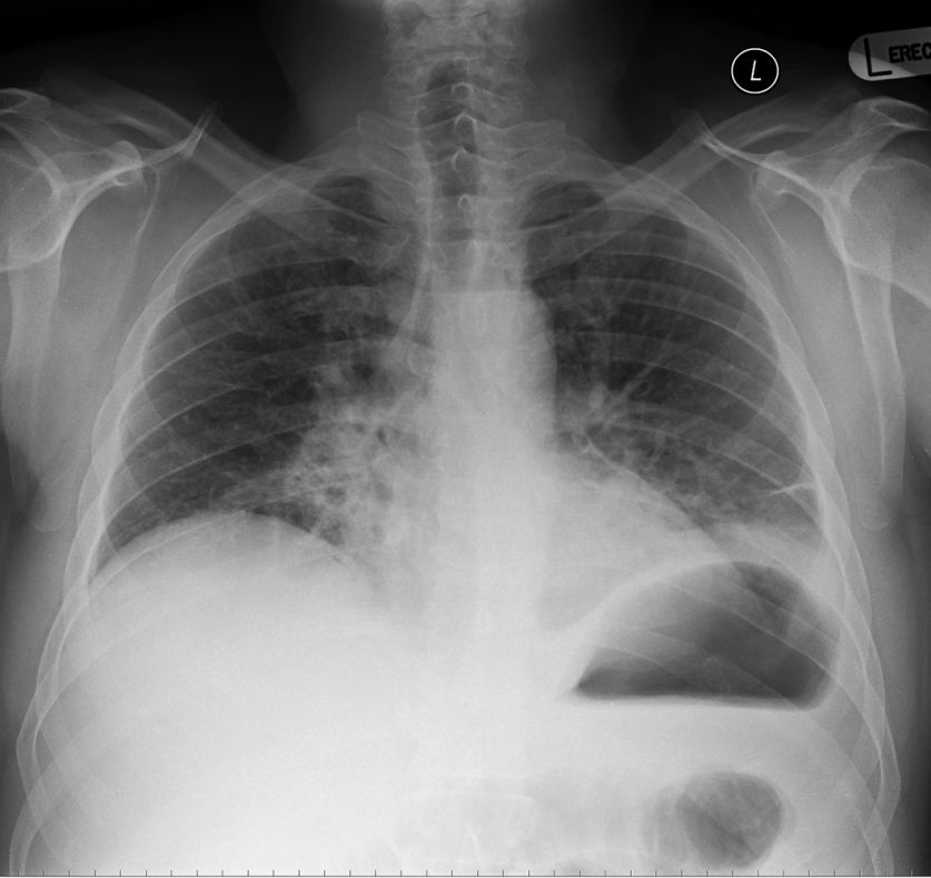
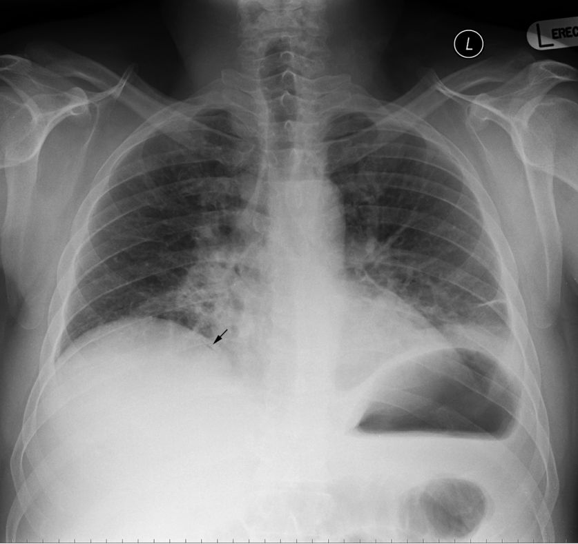
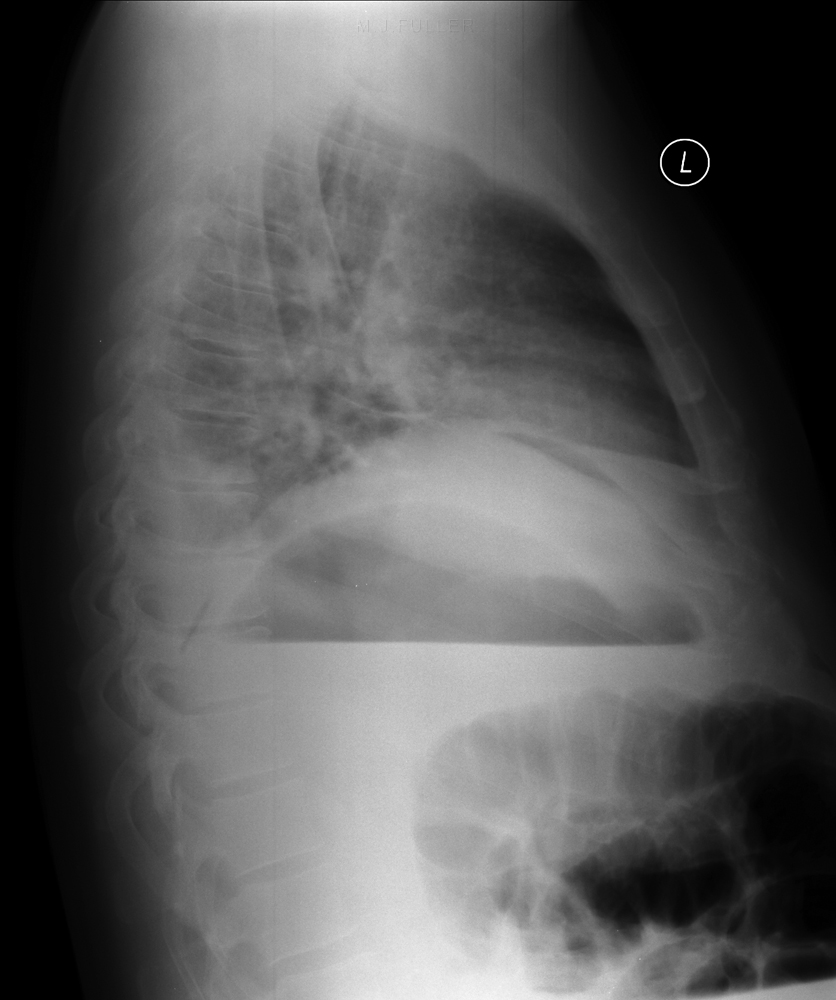
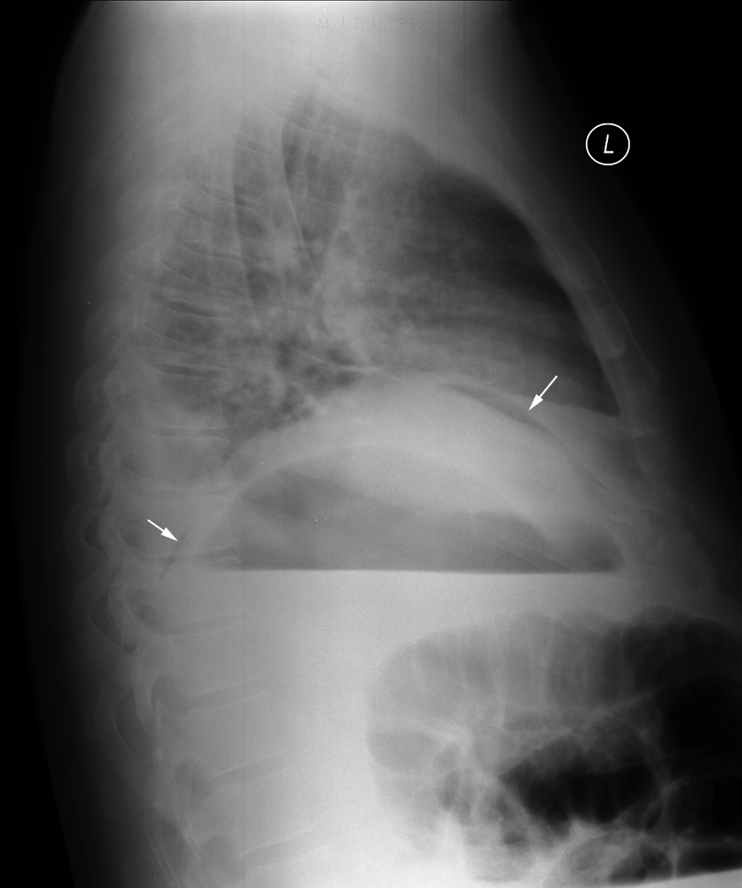
Case 1
- Pneumoperitoneum
- Pneumoperitoneum- Radiographic Techniques
- Neonatal Abdominal Pathology
- The Lateral Decubitus Abdominal Plain Film


