IntroductionThe horizontal ray lateral decubitus abdominal plain film is a legitimate alternative to the erect abdominal plain film. This page considers all aspects of this view.
Radiographic TechniqueThe lateral decubitus abdominal plain film is performed by employing a horizontal beam with the patient rolled onto their left side. This is referred to as a left lateral decubitus position. A stationary grid or wall bucky will produce superior results to a non-grid technique in adult patients. The use of an aluminium filter over the raised side will assist in providing an image of uniform density- this is particularly appropriate when using a film-screen technique.
The Right Lateral Decubitus Abdominal Plain Film
It is good practice to position the patient in a left lateral decubitus position rather than a right lateral decubitus position. The reason for this is that free intraperitoneal gas can be contrasted against the large and homogenous liver without the potentially confusing gastric fundus air. It is worth considering that if a patient is unable or unwilling to adopt the erect position or the left lateral decubitus position, there are two plain film alternatives. One is to perform a supine decubitus and the other is a right lateral decubitus.
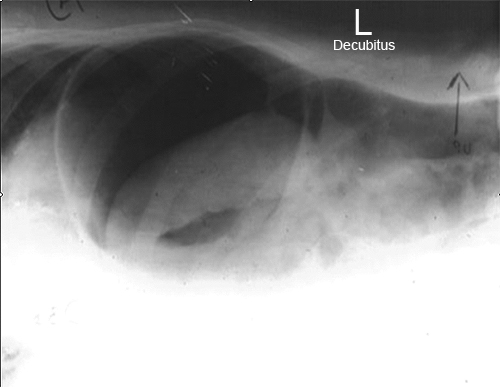
source unknown
| This image is a right lateral decubitus (right side down) which I assume was taken to demonstrate free gas. There is clear evidence of free gas. The other abdominal viscera are difficult to positively identify but could include stomach, spleen and splenic flexure.
The point of this image is that you should not be too doctrinaire in your thinking, particularly in the Emergency Department. Indeed, flexibility and working from a knowledge of first principles will often achieve the best results. |
The Supine Decubitus Abdominal Plain Film
The supine decubitus abdominal projection provides another alternative for demonstrating fluid levels and free gas.
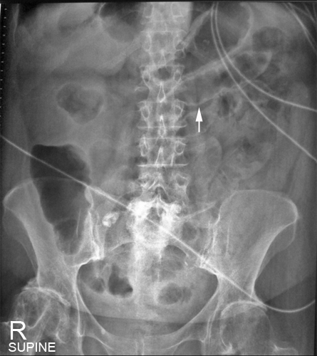
| 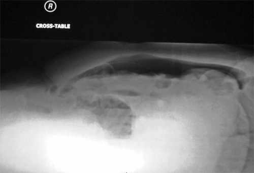 |
This patient presented to the Emergency Department with acute abdominal pain. An acute abdominal series was requested and the radiographer has performed an initial supine abdomen examination. This image revealed a Rigler's sign (arrowed) which is a positive confirmation of pneumoperitoneum. This will rarely satisfy the referring doctor in isolation- a horizontal ray projection is required. The patient was unable/unwilling to move at all. The radiographer decided to perform a horizontal ray supine decubitus abdominal film.
| The horizontal ray dorsal (supine) decubitus abdominal projection reveals free gas as expected. This projection should not be dismissed as a poor alternative to the more traditional erect and left lateral decubitus positions. Indeed, the erect lateral chest position centered on the diaphragm has been shown to be the most sensitive view for demonstrating pneumoperitoneum.
|
Case 1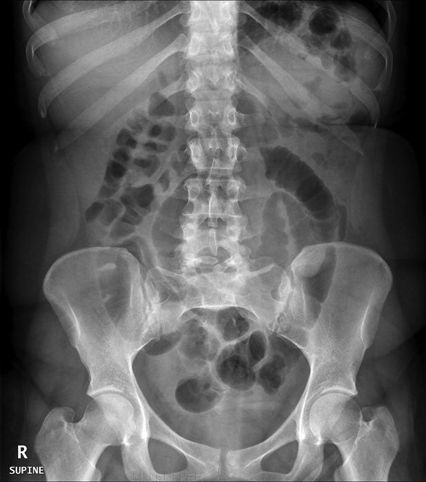 | This patient presented to the Emergency Department with abdominal pain. She was referred for abdominal plain film radiography. The supine abdominal plain film demonstrated a few prominent central gas-filled loops of small bowel. One of the gas-filled small bowel loops appeared mildly dilated. The appearances were non-specific but sufficiently concerning to warrant an erect view. |
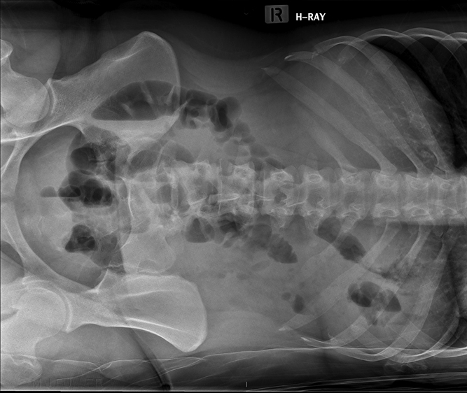 | The patient was unable to assume the erect position. The patient was transferred back onto the barouche and rolled onto her left side. The barouche was positioned in front of the erect bucky/IR and a horizontal beam was employed to produce a left lateral decubitus view. A few air/fluid levels were demonstrated. An air/fluid level in the caecum is not considered abnormal. |
The Decubitus Abdominal Techniques in the Neonatal UnitLeft Lateral Decubitus
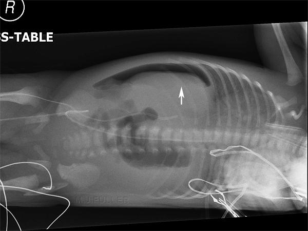 | This is the preferred decubitus position. The free intraperitoneal gas is seen easily because it is contrasted against the liver.
Although it is described as a "left lateral decubitus" it is marked with a right marker. |
Right Lateral Decubitus
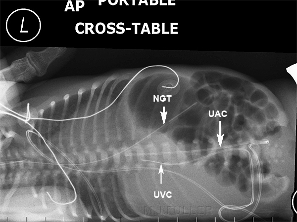 | The right lateral decubitus abdominal projection(right side down) is not the decubitus of choice. The preferred decubitus is the left lateral decubitus- left lateral decubitus is more sensitive for pneumoperitoneum. This is, nevertheless, an acceptable decubitus position when the left lateral decubitus cannot be achieved. This is also arguably preferable to a supine decubitus.
Operators phalanges noted in image. |
Supine Decubitus
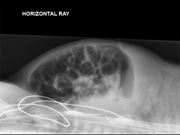 | The dorsal (supine) decubitus is a projection that can be employed when horizontal ray abdominal radiography is required in a patient that cannot be moved. This technique can be equally employed in adults, children and neonates. It is important to note that this technique is usually employed as a last resort. Despite its limitations, I would suggest that it is good radiography to adopt this view in preference to giving up.
If the referring doctor is looking for free intraperitoneal gas (pneumoperitoneum), it is important to include all of the anterior abdominal wall. Raising the baby onto a positioning sponge to improve the radiographic image is both counterproductive and of no diagnostic benefit when looking for free gas. i.e. the advantage of this technique is that you don't move the baby.
The removal of all ECG leads where appropriate will improve the image. I suspect these ECG leads were removed and placed next to the baby- arguably better to keep them out of the field altogether. |
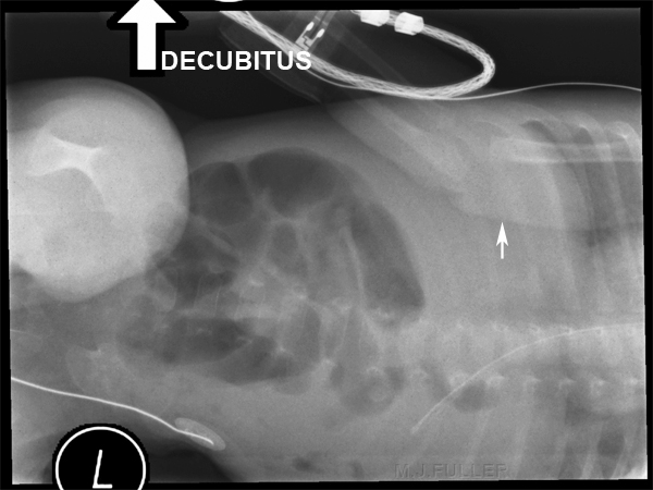 | The baby's right arm is superimposed over the chest/abdomen (white arrow). This is highly undesirable when you are looking for free gas sited at the lateral aspect of the liver |
... back to the
Wikiradiography Home page
... back to the
Applied Radiography home page


