Pneumoperitoneum- Radiographic Techniques
Pneumoperitoneum refers to free air in the peritoneal cavity. This page examines the radiographic techniques which can be employed to demonstrate free intraperitoneal gas. It is important to go to the pneumoperitoneum overview page on this Wiki before perusing this page. There are terms used on this page which are explained in the pathology overview page here.
Radiographic Techniques
“The Radiological signs of pneumoperitoneum are among the most important signs in radiology, indeed in Medicine. Sometimes the amount of free gas is small and you may have to work to demonstrate it (ie modify the film technique). Miss it and the patient may die”
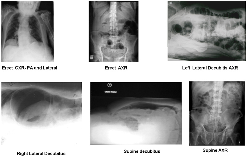
The radiographic techniques for demonstrating pneumoperitoneum are shown below (in no particular order)
The Supine Abdomen
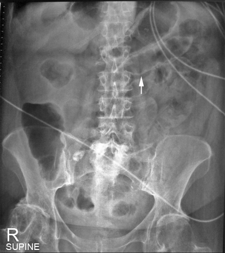
The supine abdomen is the mainstay of abdominal plain film radiography. It is most common to perform a supine abdominal examination first, even if there is an intention to follow on with other projections. It is often a careful examination of the supine abdominal image that suggests the need for further abdominal imaging.
It is important to note that the upper abdomen, including the diaphragms should be included on either the erect or the supine image.
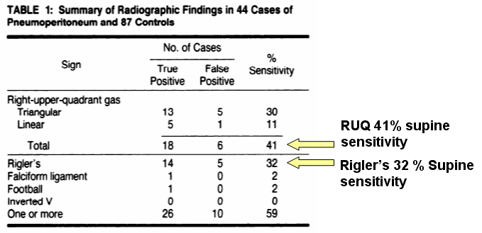
Marc S. Levine., Jac D. Scheiner. Stephen E. Rubesin Igor Laufer, Hans Herlinger.Diagnosis of Pneumoperitoneum on Supine Abdominal Radiographs.AJR 156: 731-735, April 1991You are more likely to diagnose pneumoperitoneum by close examination of the RUQ according to this study.
The Erect Abdominal Plain FilmCase Study 1a
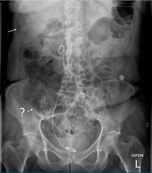
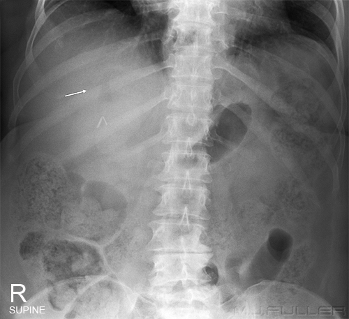
This patient presented to the Emergency Department with acute abdominal pain. The patient was referred for abdominal plain film radiography.
The radiographer has performed a supine abdominal plain film. Under close examination, it was noted that there was a linear lucency between the liver and the abdominal wall (top white arrow). There was also a question of a geographical lucency over the liver (not marked) and a possible Rigler's sign? (although this was not convincing).
There was sufficient evidence of the possibility of pneumoperitoneum to convince the radiographer to perform an erect abdominal film and erect chest examination. There was clear evidence of pneumoperitoneum on both of these images(see below).
.
The value of the erect abdominal plain film is a much debated subject (click here for my contribution to the debate). In the context of pneumoperitoneum, its value is most likely to be in the area of confirming what you already know or suspect, and in identifying other pathologies that you were not aware of.
Case Study 1b
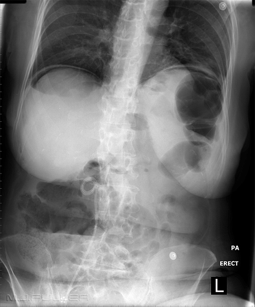
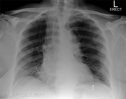
| | This is the erect plain film of the patient from case study 1a above. It is noteworthy that the patient has been positioned PA rather than AP. Most patients with acute abdominal pain are distressed and in significant discomfort. It is common for patients to prefer to "hang on" to the erect bucky/IR. They feel more secure and less likely to fall. In addition, they do not need to be repositioned for PA erect chest imaging. This image demonstrates air under both hemidiaphragms confirming the diagnosis of pneumoperitoneum. Also of note is the air-fluid level projected over the patient's liver. This suggests that the patient has hydro-pneumoperitoneum which may be of some assistance to the surgeons. The fact that the patient does not have multiple air-fluid levels may also be of interest to the surgeons. The radiographer has centred more superiorly and has managed to include much of the patient's lungfields. The high centring can be of benefit but is limited in it's utility if there is also a chest radiograph in the series. Some departments include an AXR/CXR centered on the patient's diaphragm and this is a useful projection for detecting pneumoperitoneum. |
The Left Lateral Decubitus Abdominal Plain Film
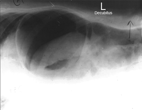
The horizontal ray lateral decubitus abdomen is frequently utilised by radiographers as an alternative to the erect abdominal plain film. This projection is particularly useful in patients who are unable to stand. The left lateral decubitus is preferred because of the potential to outline the lateral liver margin with free gas.
Case Study 2
| | This patient presented to the Emergency Department with acute abdominal pain. An acute abdominal series was requested. The radiographer considered that there was evidence of pneumoperitoneum on the supine image and proceeded to perform a horizontal ray left lateral decubitus abdominal examination. There is evidence of free air between the liver and right abdominal wall. There is also a fluid level demonstrated suggesting that the patient has a hydro-pneumoperitoneum. Importantly, there is a collection of free gas overlying the right iliac crest. This is sometimes the only site of free gas visible on the decubitus image. It is important therefore to include the pelvis on the decubitus image |
The Right Lateral Decubitus Abdominal Plain Film
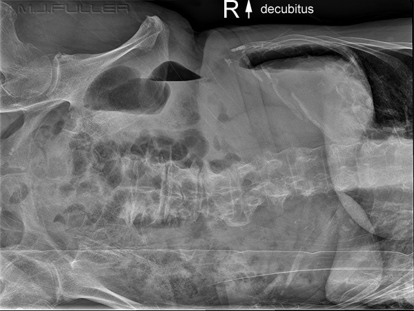
It is good practice to perform a left lateral decubitus abdominal projection rather than a right lateral decubitus abdominal projection. The reason for this is that free gas can be contrasted against the large and homogenous liver. It is worth considering that if a patient is unable or unwilling to adopt the erect position or the left lateral decubitus position, there are two plain film alternatives. One is to perform a supine decubitus and the other is a right lateral decubitus.
| | This image is a right lateral decubitus (right side down) which I assume was taken to demonstrate free gas. There is clear evidence of free gas. The other abdominal viscera are difficult to positively identify but could include stomach, spleen and splenic flexure. The point of this image is that you should not be too doctrinaire in your thinking, particularly in the Emergency Department. Indeed, flexibility and working from a knowledge of first principles will often achieve the best results. |
The Supine Decubitus Abdominal Plain Film
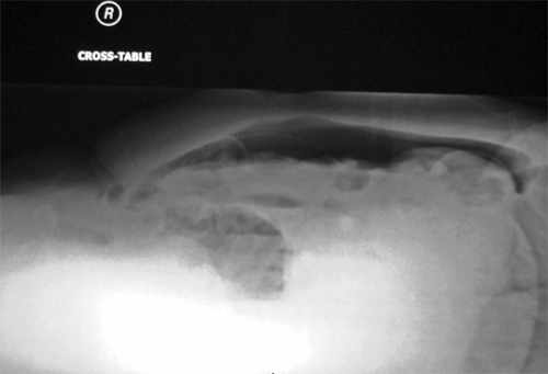
The supine decubitus abdominal projection provides another alternative for demonstrating fluid levels and free gas.
Case Study 3
The Erect Chest
The erect AP/PA chest centered on the diaphragm is widely considered the most sensitive view for detecting pneumoperitoneum. My experience and at least one research study suggest that it is the lateral chest projection that is the more sensitive. John H. Woodring and Michael J. Heiser's study that was published in the AJA compared detection rates for pneumoperitoneum in erect-PA and erect-lateral chest images and reported the following.
Projection
Detection Rate (%)
PA 80 Lateral 98 Total 100
This study reported a 98% detection rate for pneumoperitoneum on the lateral erect chest and an 80 % detection rate for the erect AP/PA chest.
Case Study 4
| |
|
| |
|
| |
|
| |
|
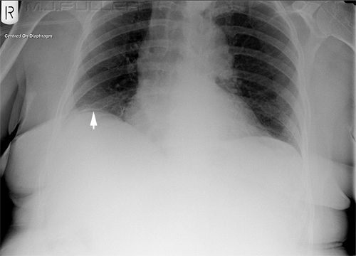
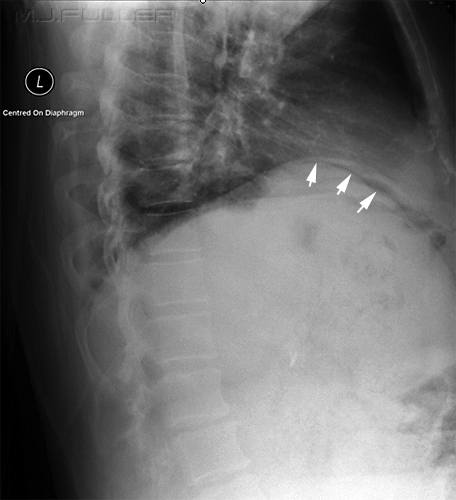
This patient proceeded to CT where there was a finding of pneumoperitoneum. It was also noted that the patient had multiple dense artifacts in the small bowel which were presumed to be bone fragments. One of these fragments appeared to be lodged in the bowel wall with localised thickening of the wall. This raised the possibility of perforation of the small bowel wall by a bone fragment.
This case illustrates a common finding in subtle pneumoperitoneum - that is, the lateral erect chest position is the most sensitive for detecting small amounts of free intraperitoneal gas. The reason for this is that free gas often collects anterior to the ventral surface of the liver. Importantly, this free gas is not evenly distributed under the dome of the right hemi-diaphragm. This is attributed to the fact that it is restricted by the peritoneal reflection at the bare area of the liver. On the erect chest image, the X-ray beam tends to just "clip" the least dependent part of the free gas collection, producing a subtle sign of pneumoperitoneum. The lateral projection however reveals the full extent of this anterior collection of free gas.
This is not simply a quaint or quirky case. This is a common scenario and is consistent with the research findings of John H. Woodring and Michael J. Heiser.
Summary
As James Begg stated "Sometimes the amount of free gas is small, and you may have to work to demonstrate it (ie modify the film technique)”.
(Abdominal X-rays made easy. 2nd edition, James D. Begg Churchill Livingstone, Elsevier, 2006p94) Pneumoperitoneum and pneumothorax pattern recognition skills are amongst the most important skills that a trauma radiographer can acquire. Together with a sound knowledge of radiographic techniques, the radiographer is in a position to play a pivotal role in the Emergency Department in detecting these pathologies.
.....back to the Applied Radiography home page
