Lateral Chest Case 11
Jump to navigation
Jump to search
Preamble
Other Relevant Wikiradiography Pages
Lateral
... back to the wikiRadiography home page
... back to the Applied Radiography home page
... back to the What is the Value of the Lateral Chest Projection? page
This is the answer page to Case 6 from the page titled What is the Value of the Lateral Chest Projection?
Other Relevant Wikiradiography Pages
- What is the Value of the Lateral Chest Projection?
- Left Upper Lobe Consolidation
- Left Upper Lobe Collapse
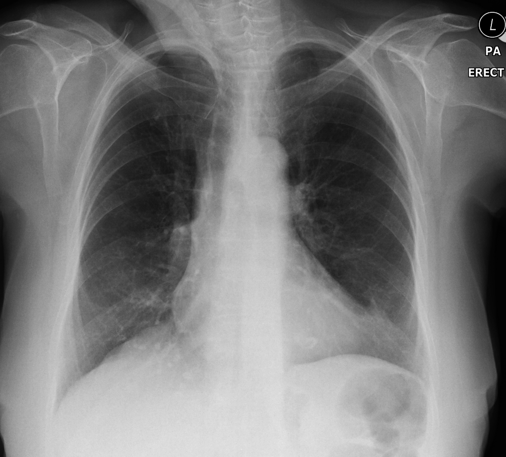
PA Erect Projection
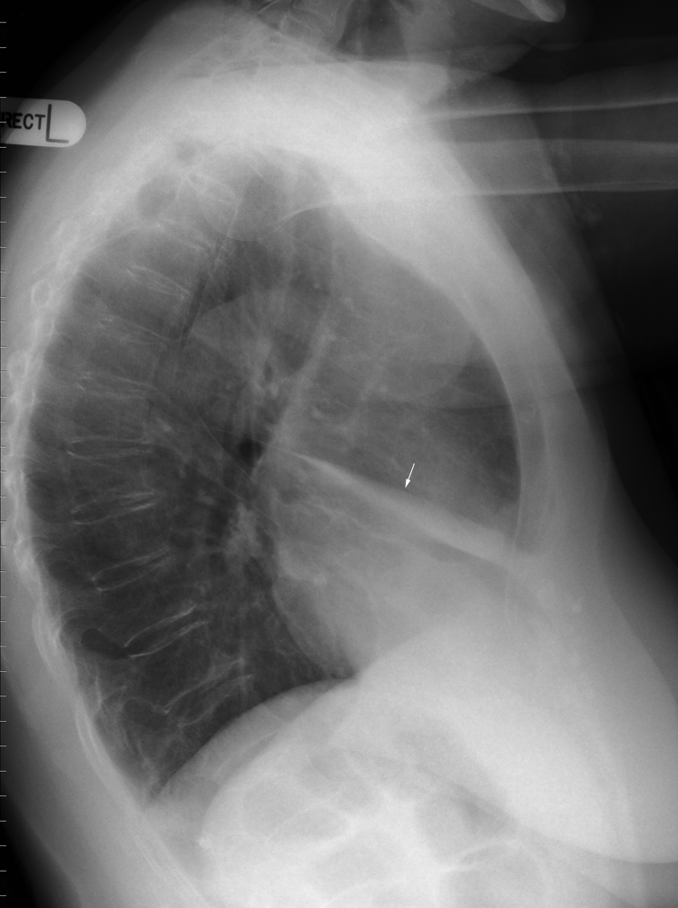
Lateral
The lateral chest demonstrates collapse/consolidation within the lingular segment of the left upper lobe.
... back to the wikiRadiography home page
... back to the Applied Radiography home page
... back to the What is the Value of the Lateral Chest Projection? page

