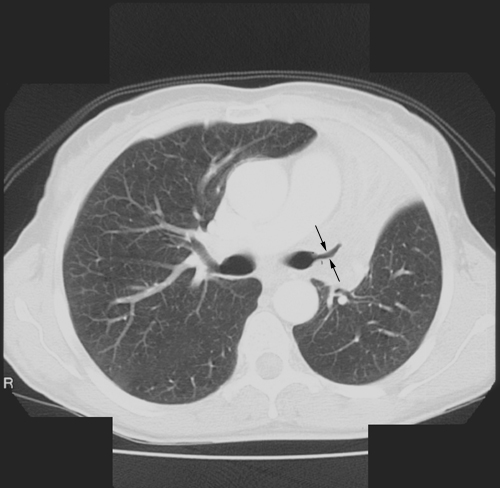
Left Upper Lobe Collapse
Jump to navigation
Jump to search
Introduction
Plain Film Signs of LUL Collapse
Plain Film Appearance
Case 1
... back to the Applied Radiography home page
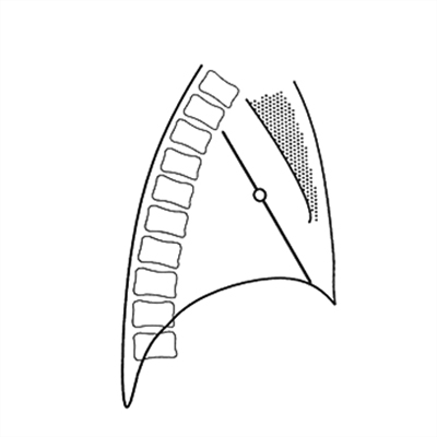
Important Characteristics of all Lobar CollapseThe left upper lobe does not collapse in the same manner as the right upper lobe. This is a legacy of anatomy. There is no middle lobe on the left- the equivalent of the RML on the left is the lingula segment of the LUL.
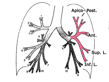
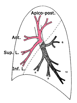
The Left Upper Lobe (LUL) Anatomy1.Collapse and consolation can occur independently or together2.Collapse can be partial or complete3.It is often not clear to what extent the appearance is due to collapse or consolidation or both. The degrees of each are often unclear.4.If a lobe is only partially collapsed and there is no accompanying consolidation, there may be no increase in opacity5.In cases of pure collapse, only when the collapse is virtually complete will there be a significant increase in density of the affected lung
adapted from <a class="external" href="http://books.google.com.au/books?id=Bif0zpmEWtAC" rel="nofollow" target="_blank">By Fred W. Wright Radiology of the Chest and Related Conditions: Together with an Extensive Illustrative Collection of Radiographs CRC Press, 2002</a>On the left there is no middle lobe; the anatomical equivalent region corresponding to the right middle lobe is known as the lingula, and like the RML, is also composed of two segments. Unlike their counterparts on the right however, the segments are stacked one on top of another, rather than side.
<a class="external" href="http://lib.cpums.edu.cn/jiepou/tupu/atlas/www.vh.org/adult/provider/radiology/LungAnatomy/RightLung/RtLungSegAnat.html" rel="nofollow" target="_blank">http://lib.cpums.edu.cn/jiepou/tupu/atlas/www.vh.org/adult/provider/radiology/LungAnatomy/RightLung/RtLungSegAnat.html</a>.
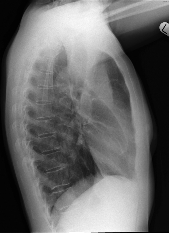
Note that upper lobe pathology could appear very low on a chest X-ray image. The upper lobe is the anterior lobe as much as it is the upper lobe.
adapted from <a class="external" href="http://books.google.com.au/books?id=Bif0zpmEWtAC" rel="nofollow" target="_blank">By Fred W. Wright Radiology of the Chest and Related Conditions: Together with an Extensive Illustrative Collection of Radiographs CRC Press, 2002</a>
More information on lung anatomy here
Plain Film Signs of LUL Collapse
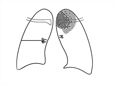
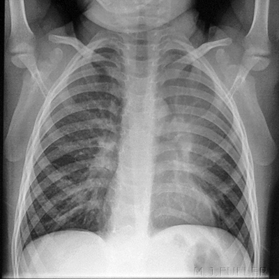
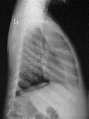
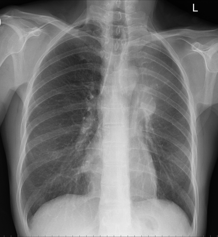
- The PA view will show an area of increased opacity in the left upper lobe with an ill-defined margin.
- The increased opacity can be very subtle and may be most evident medially.
- Unlike RUL collapse, there is no sharply defined border- the abnormal increase in lung density merges into the normal lung below.
- The incease in lung density can be almost imperceptible on the PA view. The aortic knob is often obliterated.
- Similarly, the upper left cardiac shadow can be obliterated.
- The left hilum may be elevated
- There may be a lucent stripe between the medial edge of the collpased segment and the aortic arch. The is lower lube that has been pulled up by the collpased lung (Luftsichel Sign)
Plain Film Appearance
Case 1
... back to the Applied Radiography home page
