Ulcerative Colitis and Crohn's Disease
Introduction
I have grouped these two inflammatory bowel diseases together in recognition of the possible similarities in their appearances on abdominal plain film. They are not the same disease process.
Crohn's Disease
Pathology
Crohn's disease is an inflammatory disease of the gastrointestinal tract. The causative agent has not been positively identified
Plain Film Appearances
Crohn's disease (also known as granulomatous colitis when confined to the colon) may be indistinguishable from ulcerative colitis when confined to the colon. The plain film signs of Crohn's disease are as follows:
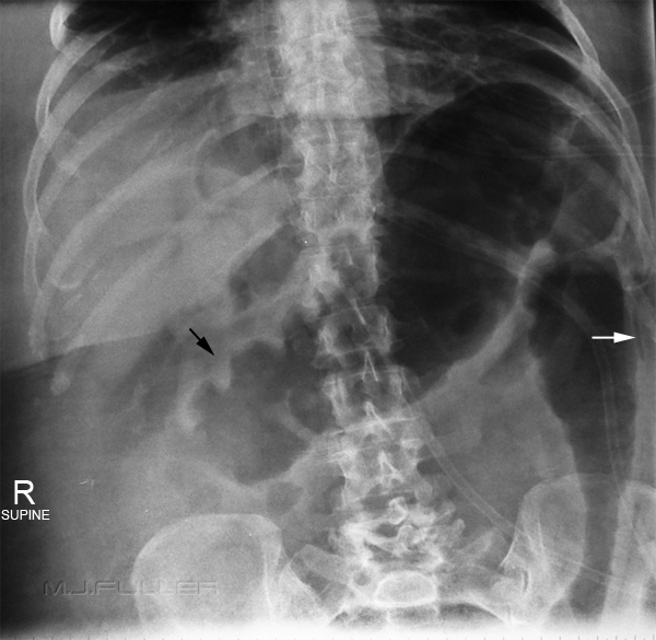
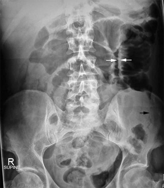
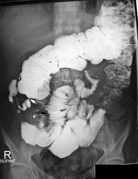
Abdominal Plain Film Signs Comment long gas-filled stricture can also be seen in other diseases including ulcerative colitis narrowed small bowel lumen not seen in ulcerative colitis small bowel obstruction indication of marked stenosis causing obstruction terminal ileum disease common site for inflammation of bowel in Crohn's disease skip lesions Crohn's lesions can be isolated lesions that occur anywhere in the GIT. Multiple isolated areas of inflammation are referred to as skip lesions bowel wall thickening sometimes seen in small bowel on plain film fistula inflammation affects whole wall thickness nd can lead to breakdown of wall and fistula formation associated with spinal arthropathy associated with sacroiliitis and ankylising spondylitis cobblestone appearance, particularly of terminal ileum seen on bariun follow-through examination intra-mural gas fulminant Crohn's disease can proceed to toxic megacolon with intra-mural gas
Patient's with fulminant Crohn's disease may be subject to daily abdominal radiographs where there is a possibility of toxic megacolon.
Case 1
Case 2
Ulcerative Colitis
Pathology
Plain Film Appearances
psedopolyps (acute fulminant stage) lumpy impressions in bowel gas, islands of lumpy mucosa absence of faeces acute and chronic stages of disease featureless, ribbon-like, lead pipe colon active and quiescent stages fine mucosal granular pattern on barium enema may be faintly visible on plain film thumbprinting (active stage) caused by mucoal oedema loss of haustration of left colon non-active stage shortening of colon ? caused by fibrosis, longitudital muscle spasm
Case 1
T


