Transthoracic Lateral Shoulder
Jump to navigation
Jump to search
Introduction
Cases
... back to the Wikiradiography home page
... back to the Applied Radiography home page
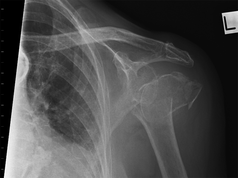
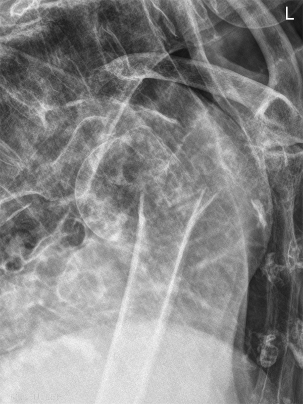
Radiographic TechniqueThe transthoracic lateral projection of the shoulder/proximal humerus is a technique that is not commonly practiced by most radiographers. The technique is a valid method for radiographic demonstration of the proximal humerus in particular and is worthy of inclusion in the radiographers' technique toolkit.
The reasons for the lack of favour of this technique are likely to be
- the inherent superimposition of the anatomy over the patient's chest
- the need for the patient to hold very still during a relatively long exposure
- and the fact that it is a very operator-dependent technique.. it takes some practice to master
<embed allowfullscreen="true" height="350" src="http://widget.wetpaintserv.us/wiki/wikiradiography/widget/youtubevideo/6d5f90a2d3200530a872a020ec1807b452d5b780" type="application/x-shockwave-flash" width="425" wmode="transparent"/> <embed allowfullscreen="true" height="350" src="http://widget.wetpaintserv.us/wiki/wikiradiography/widget/youtubevideo/8aab5aeb5984d011212a793e9831af7edfd300d4" type="application/x-shockwave-flash" width="425" wmode="transparent"/> <embed allowfullscreen="true" height="350" src="http://widget.wetpaintserv.us/wiki/wikiradiography/widget/youtubevideo/b862762ebdd2bac0d13027ec5bd3701f554af8de" type="application/x-shockwave-flash" width="425" wmode="transparent"/> These videos should provide the basic positioning technique. An exposure should be selected that provides sufficient penetration to adequately penetrate the patient's (> 70 kVp) chest and an exposure time that will provide sufficient blurring of the thoracic anatomy (at least 1 second). Shallow breathing will help to increase the blurring of the lung markings but will also increase the risk of undesirable movement unsharpness.
Notes
- Raising the unaffected arm above the patient's head will tend to cause the affected arm to drop allowing a perpendicular central ray. Alternatively, angle the beam 15 degrees cephalad.
- A breathing technique over 3 to 4 seconds will provide best results but will also increase the risk of undesirable movement unsharpness.
- If a respirated respiration technique is used, exposure on full inspiration.
Cases
... back to the Wikiradiography home page
... back to the Applied Radiography home page

