The SI Projection of the Trauma Shoulder

From wikiRadiography
Jump to navigation Jump to search
Introduction

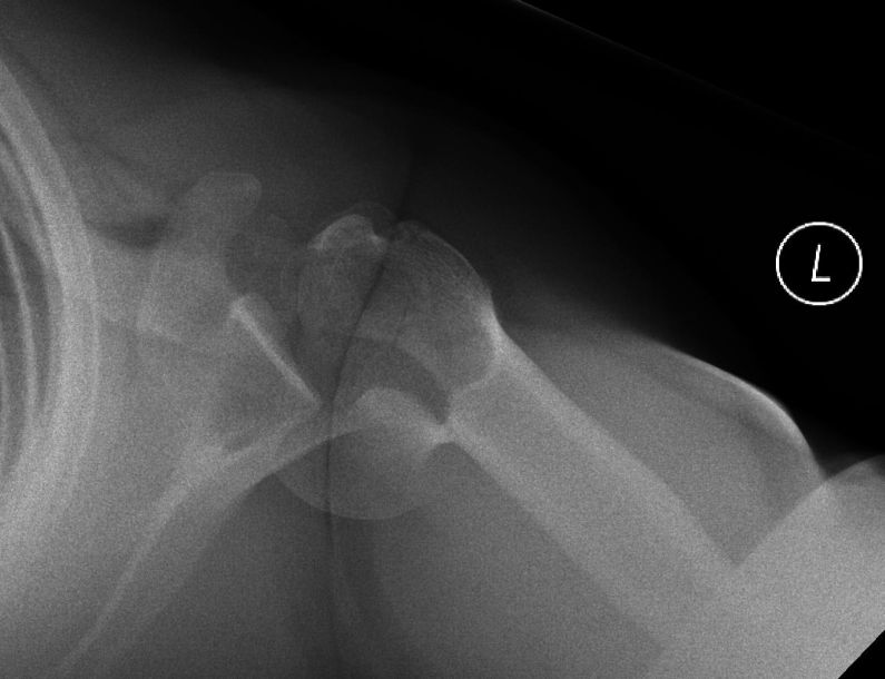
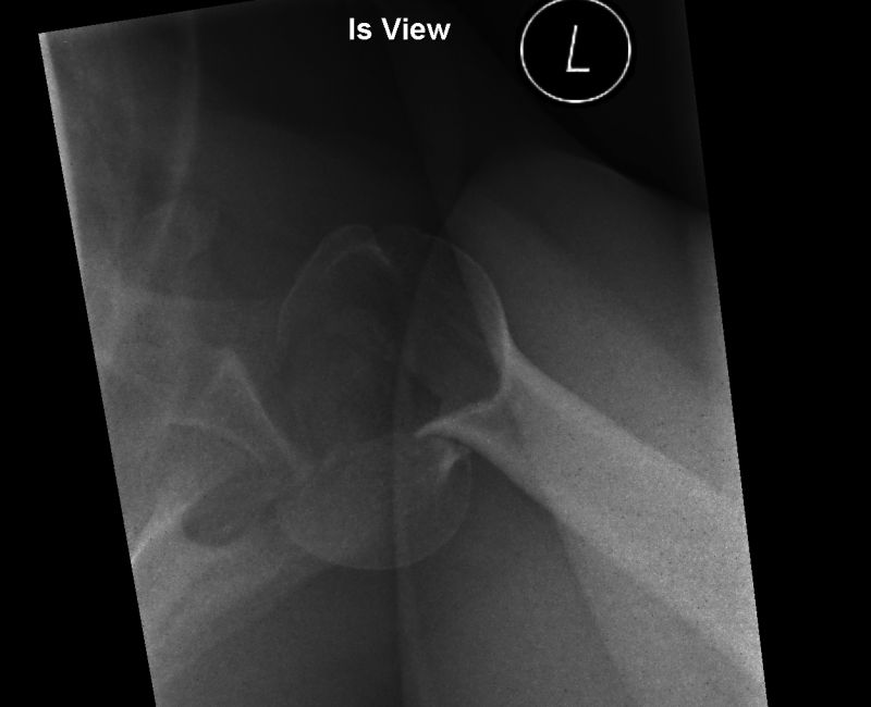
The SI/IS shoulder projection and its numerous variants often provide vital information in assessing shoulder trauma radiographically. Importantly, humeral head/neck fractures can be revealed to be fracture-dislocations after imaging the patient in the IS/SI position. It is noteworthy that an SI/IS or modified SI/IS can be performed on every shoulder trauma patient- a patient's inability to abduct his/her humerus does not preclude these projections.


Case 1
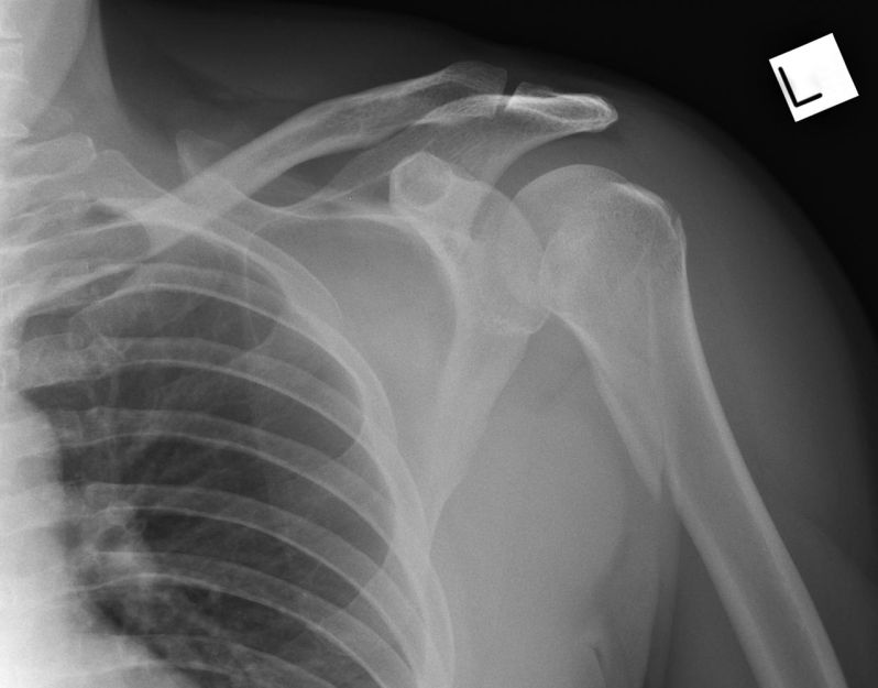
AP shoulderThis 40 year old male presented to the Emergency Department after falling off his motorbike. He was experiencing considerable shoulder pain and limitation of movement. He was referred for left shoulder radiography.

The AP shoulder projection image demonstrates a metaphyseal fracture of the proximal humerus.
SI shoulderThe SI projection image reveals a fracture/dislocation of the proximal humerus.


Case 2

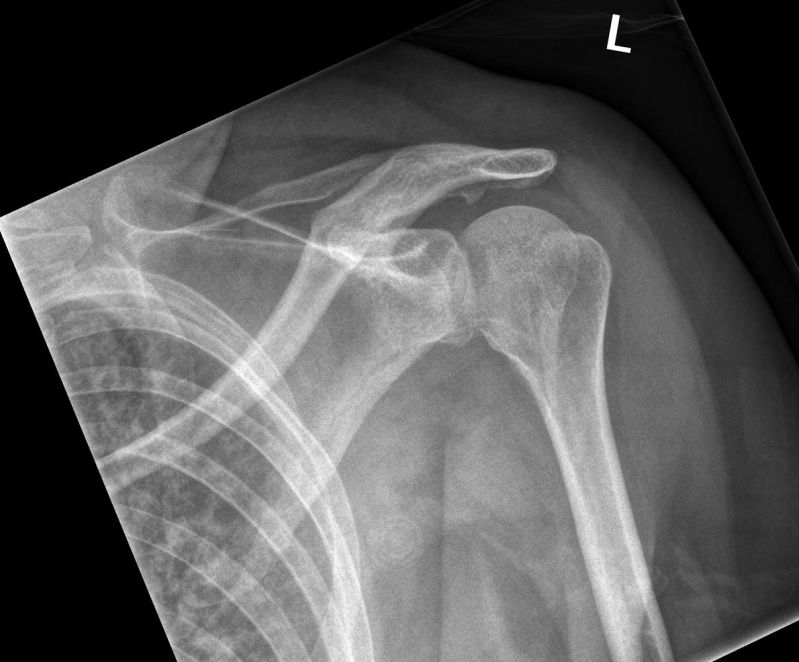
AP shoulderThis 32 year old male presented to the Emergency Department after falling off a retaining wall. He was examined and found to have a painful left shoulder with a limited range of movement. He was referred for left shoulder radiography.

Th AP shoulder projection image demonstrates an unusual appearance of the humeral head neck. There appears to to be a fracture of the humeral head/neck.

A sub- acromial spur is noted.
IS shoulderThe IS projection image reveals a fracture dislocation that was suspected on the initial AP projection image.