Tarsal Bone Fractures
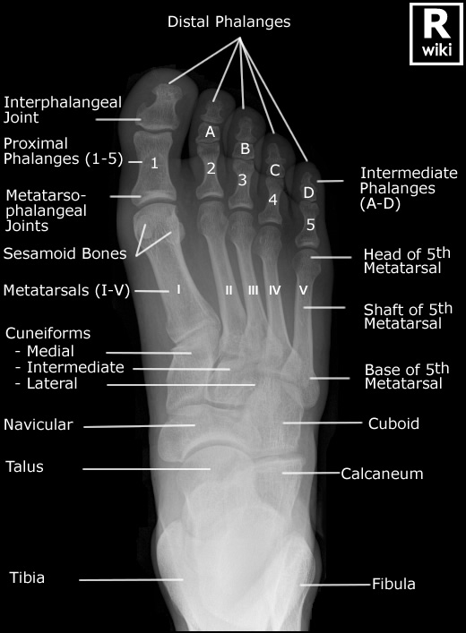
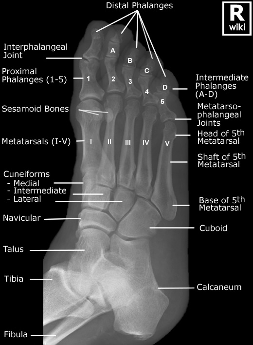
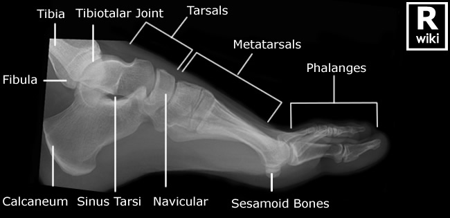
Tarsal bone fractures are less commonly seen in Emergency Department and can therefore cause difficulties in diagnosis. This page considers all aspects of the radiography of tarsal bone fractures
Acknowledgement
I have drawn extensively from the work of Nicholas Beckmann, MD and Manickam Kumaravel, MD (The Department of Diagnostic and Interventional Imaging, The University of Texas Medical School). Their presentation <a class="external" href="http://www.uth.tmc.edu/radiology/presentations/2008/nutcracker_beckman_2008.pdf" rel="nofollow" target="_blank">Nuts and Bolts of the Nutcracker</a> is recommended for further reading.
Anatomy
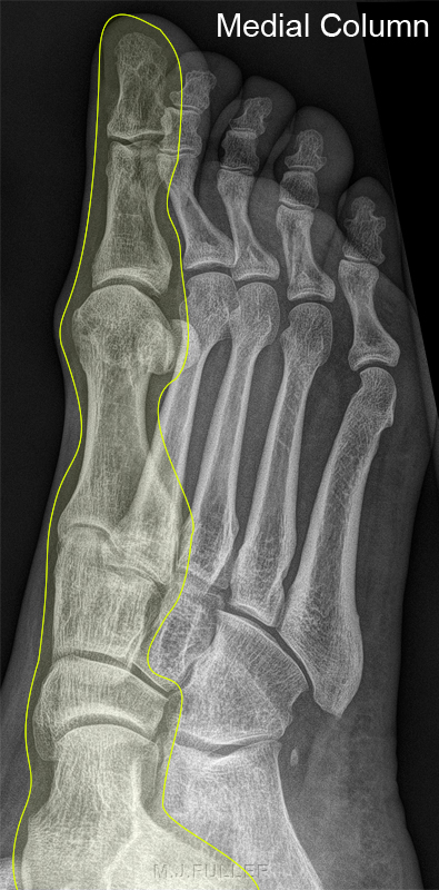
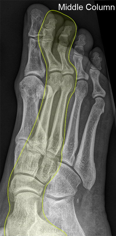
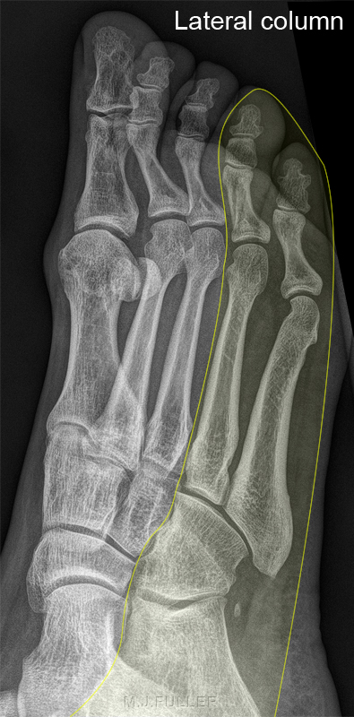
The Columns of the Foot
Medial Column of the foot •1st metatarsal
•Medial cunieform
•Navicular
•TalusLateral Column of the foot •2nd & 3rd metatarsals
•Middle & lateral cunieforms
•Navicular
•TalusLateral Column of the foot Lateral column of foot provides majority of mobility and weight bearing in the foot, Significant alterations in foot biomechanics occur with lateral column joint disruption or loss of length.•4th & 5th metatarsals
•Cuboid
•Calcaneus
Note that the cuboid articulates with the bases of the 4th and 5th metatarsals and the calcaneum.
Nicholas Beckmann, MD and Manickam Kumaravel, MD <a class="external" href="http://www.uth.tmc.edu/radiology/presentations/2008/nutcracker_beckman_2008.pdf" rel="nofollow" target="_blank">Nuts and Bolts of the Nutcracker</a> . http://www.uth.tmc.edu/radiology/presentations/2008/nutcracker_beckman_2008.pdf
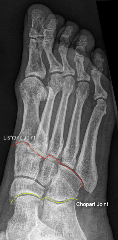
Midfoot Articlations
Midfoot articulations with forefoot and hind foot •Lisfranc joint (forefoot articulation, red line)
•Chopart joint (hind foot articulation, yellow line)
Cuboid Fractures
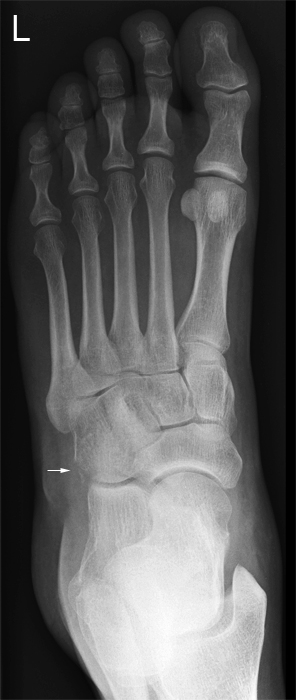
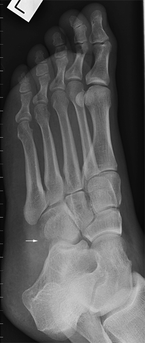
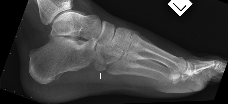
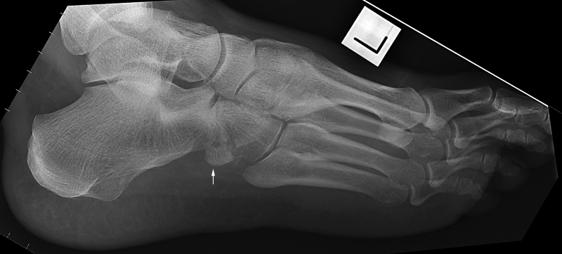
Case 1
This 50 year old male presented to the Emergency Department following a fall from a ladder. On landing his foot became stuck. He was referred for ankle radiography initially then re-referred for foot radiography.
