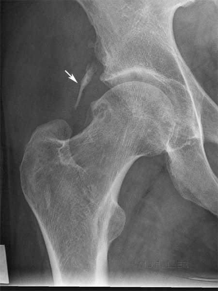
Soft Tissue Calcification
Introduction
Soft tissue calcification identification can be a useful skill for radiographers who work in the Emergency Department. Timely identification of soft tissue calcification can expedite the patient's imaging and save the patient from unnecessary additional imaging (particularly comparison views). From the Radiologist's point of view, soft tissues calcifications are usually not problematic, but occasionally pop up as incidental findings and, as one author noted, "it behoves the radiologist to say something intelligent about them" <a class="external" href="http://www.rad.washington.edu/academics/academic-sections/msk/teaching-materials/online-musculoskeletal-radiology-book/soft-tissue-calcifications" rel="nofollow" target="_blank">http://www.rad.washington.edu/academics/academic-sections/msk/teaching-materials/online-musculoskeletal-radiology-book/soft-tissue-calcifications
</a>
Types of soft tissue calcification
Dystrophic Calcification
Metastatic Calcification
- Calcinosis
- Chondrocalcinosis
- Synovial Chondromatosis.
Type Incidence Examples Appearance Dystrophic Calcification 95 - 98 % small to large amorphous Ca++ in the damaged tissue -- may progress to ossification (formation of cortex and medullary space are then seen) Metastatic Calcification 1 - 2% Entities such as renal failure, hyperparathyroidism, sarcoidosis, milk-alkali syndrome, etc. can lead to metastatic calcifications. finely speckled calcification throughout soft tissues Calcinosis 1 - 2% Calcification of cutaneous, subcutaneous or deep connective tissue.
- Not associated with metabolic disturbance.
- May be associated with collagen-vascular disease.
- 3 Types:
1. Calcinosis circumscripta
2. Calcinosis universalis
3. Tumoral calcinosisChondrocalcinosis
(Calcium pyrophosphate dihydrate deposition disease- CPPD)1 - 2 % Non-specific calcification of cartilage
Gout -- calcium urate crystals
Pseudogout - calcium pyrophosphate
Ochronosis – homogentisic acid oxidase
Hyperparathyroidism
Diabetes
Degenerative disease
- (CPPD) is usually associated with chondrocalcinosis.
This typically appears as a fine white line overlying the hyaline articular cartilage.- CPPD is also associated with calcifications in the soft tissues of the spine.
Synovial Chondromatosis Synovial chondromatosis
Affects knee, hip and shoulder joint.
Tends to be mono-articular.
Due to metaplasia of synovial connective tissue.
Uncommon cause of loose bodies.
Biopsy shows active synovial proliferation
<a class="external" href="http://www.rad.washington.edu/academics/academic-sections/msk/teaching-materials/online-musculoskeletal-radiology-book/soft-tissue-calcifications" rel="nofollow" target="_blank">http://www.rad.washington.edu/academics/academic-sections/msk/teaching-materials/online-musculoskeletal-radiology-book/soft-tissue-calcifications</a>
The Department of Radiology at the University of Washington provide a simple english description of dystrophic calcification as follows
| "As you can see, almost every calcification that one sees in the soft tissues in actual radiographic practice is due to dystrophic calcification. What does this mean? Simply this: when tissue is damaged, the body responds to this injury in a nonspecific manner by invoking the generic inflammatory response reaction. This sometimes ends with calcification of the damaged tissue. This calcification is probably usually only microscopic, but is occasionally enough to be seen radiographically." | <a class="external" href="http://www.rad.washington.edu/academics/academic-sections/msk/teaching-materials/online-musculoskeletal-radiology-book/soft-tissue-calcifications" rel="nofollow" target="_blank">http://www.rad.washington.edu/academics/academic-sections/msk/teaching-materials/online-musculoskeletal-radiology-book/soft-tissue-calcifications</a> |
- Calcification in damaged or degenerating tissue by small to large amorphous calcium deposit.
- Not associated with metabolic disorder.
- Almost every calcification that one sees in the soft tissues in actual radiographic practice is due to dystrophic calcification.
- They have a prevalence of 95-98% out of all soft tissue calcification.
Differential diagnosis of dystrophic calcificationMnemonic = VINDICATE (VINDAT)
V = Venous - Phlebolith
I = Infection - Cysticercosis
N = Neoplasm - Osteosarcoma
D = Drugs - Vitamin D overdose
A = Autoimmune - Dermatomyositis
T = Trauma - Hematoma, Heterotopic ossification
2. Metastatic Calcification
... back to the Wikiradiography home pageMetastatic calcification is defined as the deposition of calcium salts in previously normal tissue due to abnormal biochemistry with disturbances in the calcium or phosphorus metabolism. (Grech P, Martin TJ, Barrington NA, Ell PJ. Diagnosis in metabolic bone disease. London: Chapman and Hall Medical; 1985:194-241.) "It occurs as opposed to dystrophic calcification where blood levels of calcium are normal, and abnormalities or degeneration of tissues result in mineral deposition. These differences in pathology also mean that metastatic calcification is often found in many tissues throughout a person or animal, while dystrophic calcification may be localized." <a class="external" href="http://en.wikipedia.org/wiki/Metastatic_calcification" rel="nofollow" target="_blank">(http://en.wikipedia.org/wiki/Metastatic_calcification)</a>. We tend to associate the term metastatic to the spread of a cancerous tumour- the term metastatic is a broader term referring to the spread of disease from one part of the body to another (not just metastatic cancer).
"The exact pathophysiology of the calcifying mechanism in metastatic calcification is not well understood, although it is thought to relate to serum calcium and phosphorus concentrations. Tissues comprising an alkaline environment are more susceptible to calcification, and therefore common sites of calcification include the lungs, gastric mucosa, and kidneys. This condition contrasts with that of dystrophic calcification, in which deposits occur in damaged tissue in the presence of normal calcium metabolism." <a class="external" href="http://www.hkcr.org/publ/Journal/vol5no3/full/Vol.5+No.+3_p.186-189.pdf" rel="nofollow" target="_blank">(KF Tam, K Wang, WC Fan. Metastatic Calcification, J HK Coll Radiol 2002;5: p 187)</a>
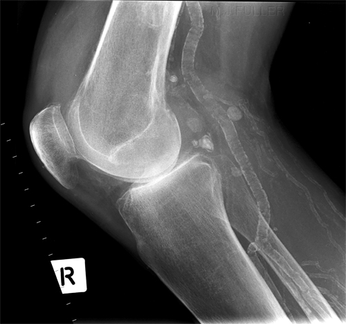
Renal Failure
This is a 'rolled' lateral knee in a patient with end stage renal failure (ESRF). There is extensive metastatic calcification involving the popliteal and tibial arteries.
...back to the Applied Radiography home page

