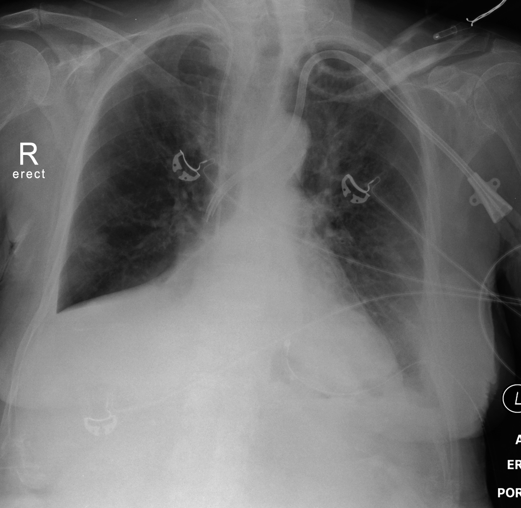
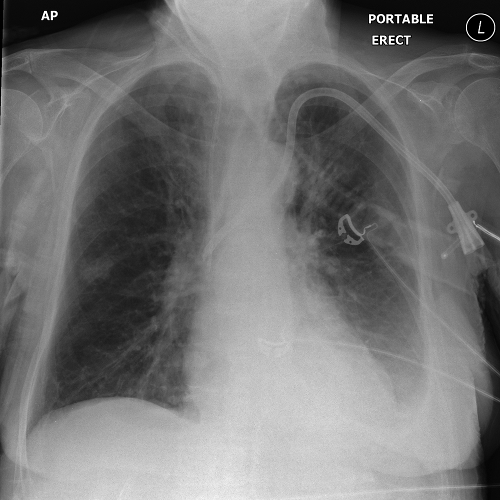
Right Lower Lobe Collapse
Jump to navigation
Jump to search
Introduction
Plain Film Appearance
Horizontal Fissure or Oblique Fissure?
Combined RML and RLL collapse
Case 1
Case 2
... back to the Applied Radiography home page
Important Characteristics of all Lobar CollapseRight lower lobe collapse is relatively uncommon. THis page considers all aspects of the plain film appearance of right lower lobe collapse.
The Right Lower Lobe Anatomy1. Collapse and consolation can occur independently or together2. Collapse can be partial or complete3. It is often not clear to what extent the appearance is due to collapse or consolidation or both. The degrees of each are often unclear.4. If a lobe is only partially collapsed and there is no accompanying consolidation, there may be no increase in opacity5. In cases of pure collapse, only when the collapse is virtually complete will there be a significant increase in density of the affected lung
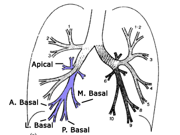
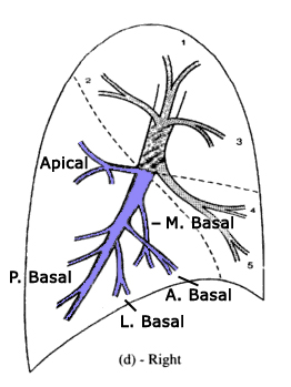
adapted from <a class="external" href="http://books.google.com.au/books?id=Bif0zpmEWtAC" rel="nofollow" target="_blank">By Fred W. Wright Radiology of the Chest and Related Conditions: Together with an Extensive Illustrative Collection of Radiographs CRC Press, 2002</a>The right lower lobe is comprised of five pulmonary segments. It is a large lobe and will provide varying patterns of collapse depending on which segments are involved.
adapted from <a class="external" href="http://books.google.com.au/books?id=Bif0zpmEWtAC" rel="nofollow" target="_blank">By Fred W. Wright Radiology of the Chest and Related Conditions: Together with an Extensive Illustrative Collection of Radiographs CRC Press, 2002</a>Note that collapse of the apical segment will not result in loss of the diaphragmatic outline.
Further information on lung anatomy here
Plain Film Appearance
- The Right lower Lobe (RLL) collapses in a manner that is similar to closing a fan.
- May mimic a RML collapse if the superior segment of the RLL is affected in isolation
- may be loss of the visualisation of the right hemidiaphragm
- There should be no loss of visualisation of the right heart border
- There may be compensatory hyperexpansion of the RML resulting a clearly dilineated right heart border and right hemidiaphragm
- There may be evidence of reduced overall lung volume on the right
- the right hilum may be displaced inferiorly (but may be obscured by the collpased lung)
- The mediastinal vessels and fat can move to the right producing a triangle of opacity to the right of the trachea (superior triangle sign)
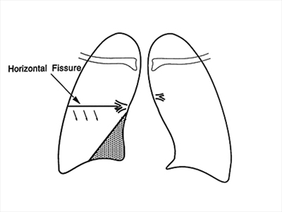
- The collapsed oblique fissure can become visible on the AP image mimicking the horizontal fissure
- The PA view will show an area of opacity at the base of the right lung adjacent to the right heart border
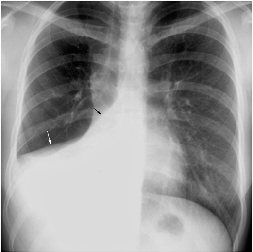
adapted from Lieberman's <a class="external" href="http://eradiology.bidmc.harvard.edu/Classics/item.aspx?section=Infectious+Diseases&labelpk=459693aa-bd69-452b-b55e-5c46b71aa9cc&pk=8a8fb39f-70e3-4739-a4f6-71466a785a26" rel="nofollow" target="_blank">Classics Collection in Radiology</a>
- The oblique fissure is displaced inferiorly (mimicking the horizontal fissure)
- There is increased density adjacent to the right heart border but not obliterating the right heart border
- There is loss of visualisation of the right hemidiaphragm (silhouette sign)
- The right hilum is displaced inferiorly
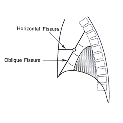
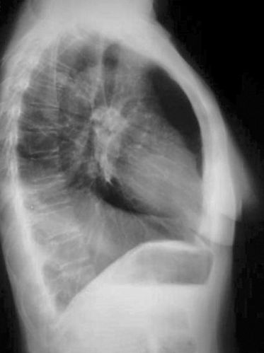
- The lateral view is usually definitive- there will be postero-inferior movement of the oblique fissure whilst maintaining the same slope
- Oblique fissue may be straight or concave anteriorly (opposite to the shape shown in the graphic!)
adapted from Lieberman's <a class="external" href="http://eradiology.bidmc.harvard.edu/Classics/item.aspx?section=Infectious+Diseases&labelpk=459693aa-bd69-452b-b55e-5c46b71aa9cc&pk=8a8fb39f-70e3-4739-a4f6-71466a785a26" rel="nofollow" target="_blank">Classics Collection in Radiology</a>
- The oblique fissure is displaced posteriorly and inferiorly
- There is loss of the normal darkening of the thoracic vertebral bodies inferiorly
- There is loss of visualisation of the right hemidiaphragm
Horizontal Fissure or Oblique Fissure?
Combined RML and RLL collapse
Case 1
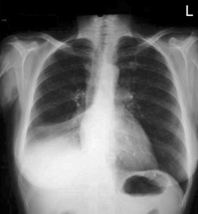
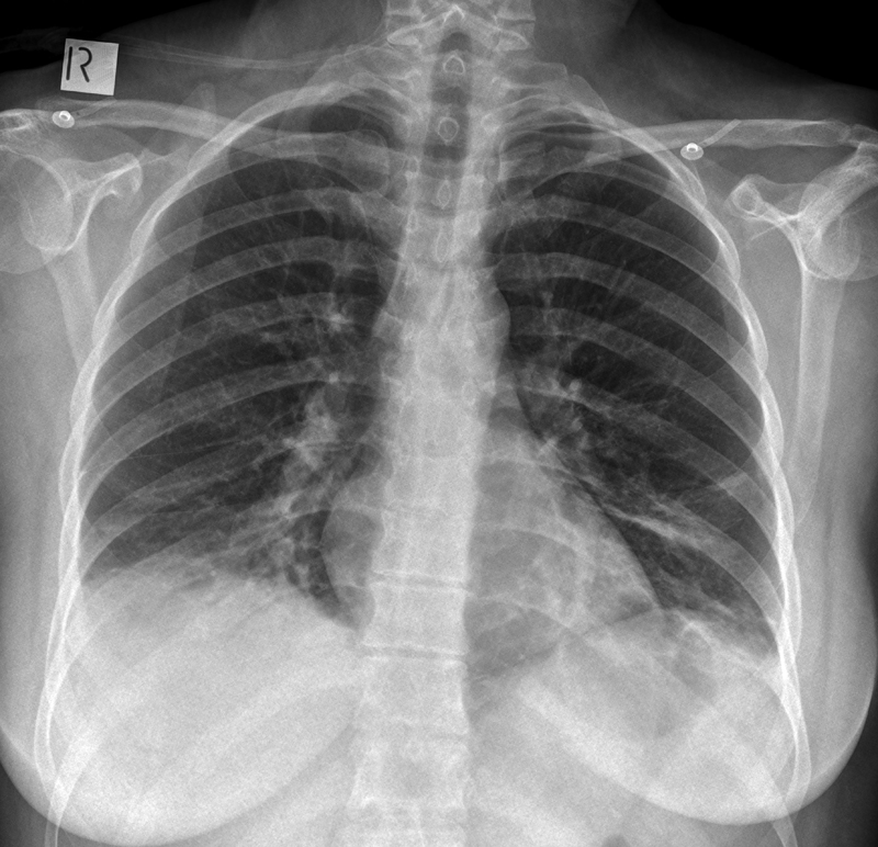
This 39 year old lady presented to the Emergency Department with shortness of breath. On auscultation she was noted to have decreased air entry on the right side. She was referred for chest radiography.
There is loss of clarity of the right hemidiaphragm. There is depression of the horizontal fissure with a faintly visible second fissure below the horizontal fissure.
There is a suggestion of increasd density of the right subdiahragmatic region compared to the corresponding subdiaphragmatic area on the left
Left linear subsegmental atelectasis noted on the left.<img align="bottom" alt="RLL Collapse" src="http://image.wikifoundry.com/image/1/s_Uv7tr4SDYKGWQ3B3eglg235206" title="RLL Collapse"/> The horizontal fissure is faintly visible.
The oblique fissure has moved infero-posteriorly indicating RLL collapse. The fissure is "S" shaped which may explain the appearence of a second fissure below the horizontal fissure on the AP/PA view.
The posterior aspect of the right hemidiaphragm is obliterated (silhouette sign)
The RLL is not showing signs of complete collpase- the collapse segments appear to be the basal segments.
Diagnosis- Partial RLL collapse
Case 2
... back to the Applied Radiography home page
