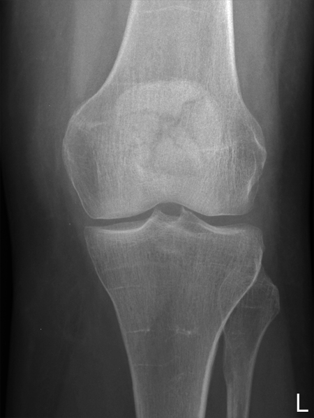
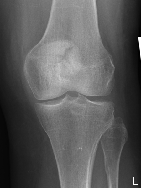
Radiography of Sesamoid Bones
Introduction
What is a sesamoid Bone?Apart from patellar radiography, specific radiography of sesamoid bones is uncommon. This page considers all aspects of radiography of sesamoid bones.
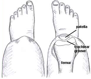
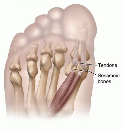
Sesamoid Bone PathologyA sesamoid bone is a bone embedded within a tendon. Sesamoid bones are found in locations where a tendon passes over a joint, such as the hand, knee, and foot. Functionally, they act to protect the tendon and to increase its mechanical effect. The presence of the sesamoid bone holds the tendon slightly farther away from the center of the joint and thus increases its moment arm. Sesamoid bones also prevent the tendon from flattening into the joint as tension increases and therefore also maintain a more consistent moment arm through a variety of possible tendon loads. <a class="external" href="http://en.wikipedia.org/wiki/Sesamoid_bone" rel="nofollow" target="_blank">http://en.wikipedia.org/wiki/Sesamoid_bone</a> The largest and most commonly imaged sesamoid bone is the patella.
Radiography of Sesamoid BonesSesamoiditis and sesamoid fracture are the most common pathologies involving sesamoid bones. Other sesamoid bone pathologies include arthritis and osteochondritis.
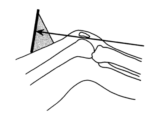
Sesamoid Bones Tendon Anatomy Radiography Patella Quadricepts tendon Skyline View
Sunrise View
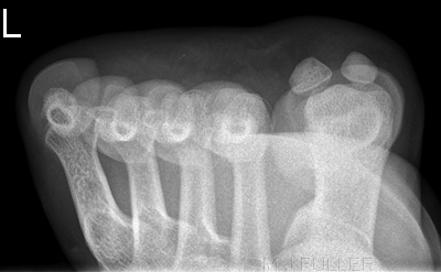
Merchant's View1st MP Joint Foot Flexor Hallicus Longus
PA vs AP Patella
