Radiography of VP Shunts
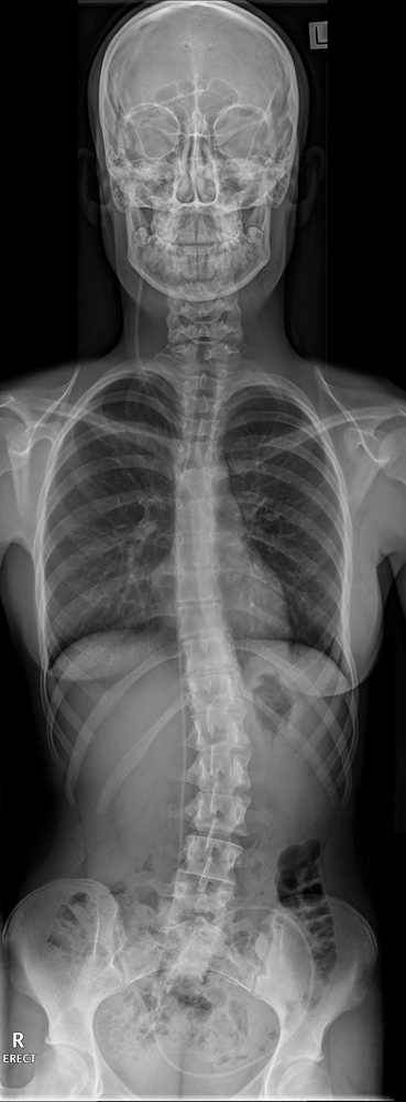
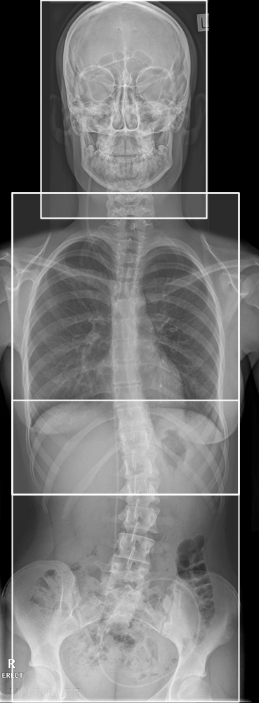
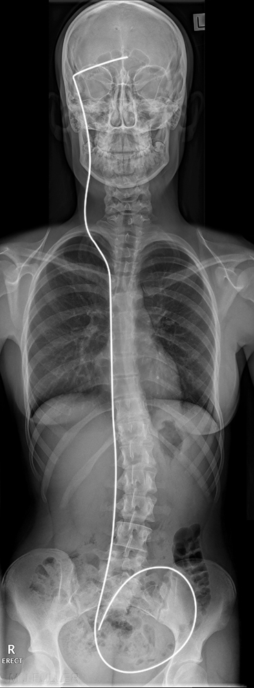
Radiography of a VP shunt is essentially a simple radiographic series. A knowledge of the course of the shunt and common shunt abnormalities will assist the radiographer in providing an appropriate set of images. Some institutions will include lateral and tangential shunt valve projections.
What is a VP Shunt?
The Ventriculo-Peritoneal (VP) shunt is a thin tube that is placed inside the brain’s ventricle and tunneled underneath the skin to the peritoneum. The peritoneum is a membrane that lines and protects the abdominal cavity and its contents. The purpose of the VP shunt is to reduce the pressure of cerebral spinal fluid (CSF) in the brain by draining it into the abdominal (peritoneal) space. The shunt tubing does not come into direct contact with the stomach or other organs. The peritoneum is the usual, safe site to place the end of the ventricular shunt, however, certain circumstances may require placing the end of the shunt tubing into a blood vessel leading to the heart (Ventriclo-Vascular or Atrial, VA) or into the pleural space of the chest (Ventriculo-Pleural, VP). <a class="external" href="http://www.divideclassic.org/Documents/ShuntInfo.htm" rel="nofollow" target="_blank">[adapted from Department of Neurology, UCSF, 1986, in Janet Freeman and Jennifer Diabato, HYDROCEPHALUS INFORMATION SHEET]</a>. Usually VP shunts are placed to treat hydrocephalus (hydro = water, cephalus = head). Many different diseases block the flow of CSF through the brain, including: brain tumours, subarachnoid hemorrhage, meningitis, and head injury. Many cases of hydrocephalus are congenital - they are caused by improper development of the CSF pathways. Shunts are not perfect, they may malfunction, clog, or break. One problem with shunts has been infection. Shunts are made of foreign material, and thus are more prone to infection than normal body tissue. Many studies suggest that shunt infections are due to microscopic contamination at the time of implantation. Shunt infection rates in the 1990's were often reported to be in the 5-9% range, far higher than for other implanted foreign bodies, such as pacemakers or joint replacements. Following VP stent insertion, a follow up CT (or MR) is sometimes obtained to verify that the ventricular catheter tip is located at the desired location in the anterior horn of the lateral ventricle close to the foramen of Monro. <a class="external" href="http://www.pauldurso.com/surgery_cranial_VP_Shunt.htm" rel="nofollow" target="_blank">(Paul S. D'Urso)</a>
VP Shunt Videos
<embed allowfullscreen="true" height="350" src="http://widget.wetpaintserv.us/wiki/wikiradiography/widget/youtubevideo/40646cc1bcf6494cfdc6c486c7880a9d4f84c796" type="application/x-shockwave-flash" width="425" wmode="transparent"/>
VP Shunt Radiography
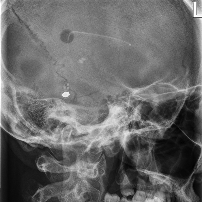
Shunt Valve Radiography
There are many different kinds of ventricular shunts. All have three basic parts: a To assess the shunt valve a dedicated shunt position is required. This is discussed further here
- Ventricular Catheter
- Shunt Valve
- Distal Catheter.


