This page contains podiatry X-ray positioning
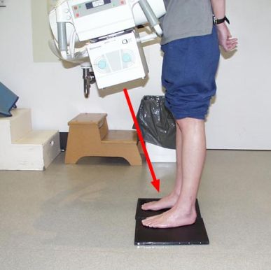
----------- work in progress -------------- Weight Bearing Dorsoplantar (DP) view
Dorso-plantar projection or Anterior-Posterior (AP) weightbearing
The patient stands erect on the cassette in the normal and base of gait
Use a single 10x12 for both feet - two on one
Central Ray - Directed at base of 3rd metatarsal
- 20-25° posteriorly (towards the heel)-anything less will get tube pushing against pt.
FFD Bones visualised - Phalanges, metatarsals, navicular, cuneiforms and cuboid
Joints visualised - Midtarsal Joint, Lisfranc Jointt, Metatarsal Phalageal Jointst, Interphalangeal Joints
| |
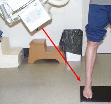
Medial Oblique View
The patient stands erect on the cassette in the normal and base of gait
make sure pt is standing - foot of interest on a 10x12 and the other foot just next to it to evenly distribute weight.
Central Ray - Directed at base of the 3rd metatarsal
- Tube angled at 35 degrees
FFD Bones visualised - Cuboid and navicular are clearly seen, but the cuneiforms are superimposed upon each other. All other bones distal to the midfoot are clearly visualised
Joints visualised: - Talonavicular, navicularcuneiform, cuboidnavicular, cuboid-lateral cuneiform, calcaneocuboid, Lisfranc's joint. All Metatarsal phalangeal Joints
|  |
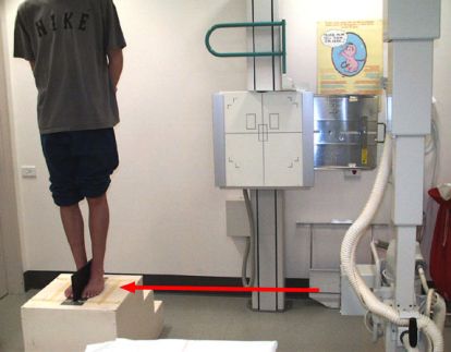
Lateral Weightbearing View
The patient stands on an Orthoposer box, that elevates the foot enough so that the X-ray beam can be angled 90° from vertical, and the central ray directed to the base of the metatarsals. The cassette is inserted into a slot in the orthoposer to hold it in position. - no devise like this one here, laterals shot medial-lateral instead, with cassette up against bucky.
Central Ray - Centered to the base of the metatarsals
- Perpendicular to IR
FFD Bones visualised
- talus, calcaneus, cuboid, navicular, medial cuneiform, 1st metatarsal.
Joints visualised:
- Midtarsal Joint, Subtalar Joint, 1st metatarsal-cuneiform, navicular-1st cuneiform.
|  |
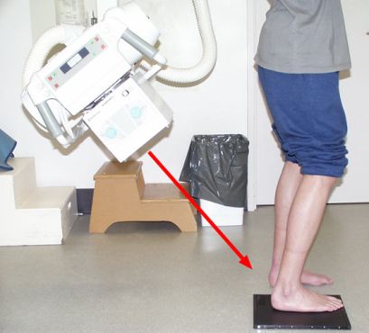
Axial calcaneal weightbearing
Also known as Harris and Beath (Coalition View)
The patient stands erect on the cassette in the normal and base of gait
DR LATT - will order these, always do both heels. Have pt bend knee to get in almost a squat position-open up collimation you must see light over calf - this will give them the tib/fib shot they need to measure - ALSO CALLED ALIGNMENT VIEW.
Central Ray
- Central beam angled 45 deg toward the midline of the heel
FFD Bones visualised
- Medial and Lateral malleoli, talus, calcaneus
Joint visualised
- Subtalar Joint, ankle Joint
|  |
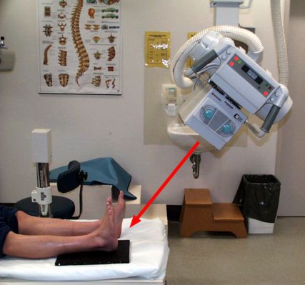
Axial calcaneal
The patient lays supine on the X-ray table with the cassette under the heel, have pt pull back toes as much possible with a bandage strip - this will get the joint visualized.
Central Ray
- Central beam angled 45 deg toward the midline of the heel
FFD
Bones visualised
- Plantar-posterior aspect of the calcaneus
Joints visualised
|  |
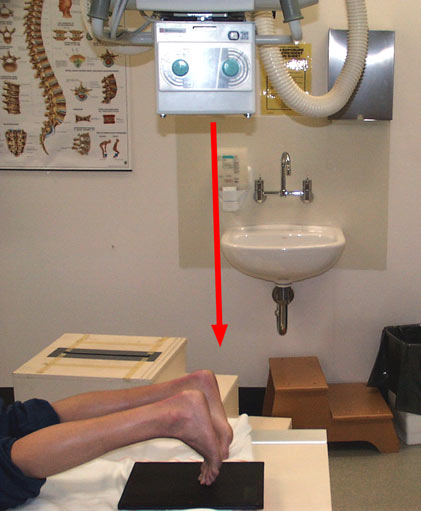
Axial sesamoid nonweightbearing
Patient is positioned prone on the X-ray table with the cassette placed beneath the foot
Central Ray
- Central ray aimed at the midline of the foot.
FFD Bones visualised
- Sesamoids and Sagittal plane relationship of metatarsal heads
Joint visualised
|  |
...Go back to Podiatry Homepage