Clinical Presentation
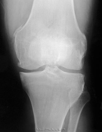
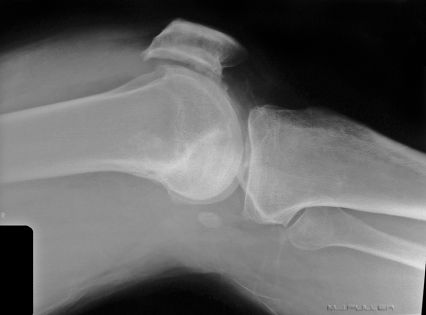
This patient presented to the Emergency Department with a painful distended left knee. A knee x-ray examination was performed and the images are shown above. What abnormal findings are demonstrated?
Images
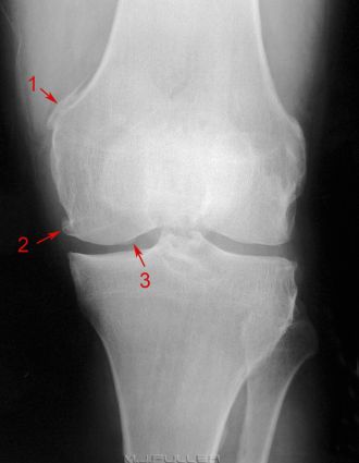
Findings  | There is abnormal bone growth adjacent to the medial condyle of the knee on the AP view (arrow 1). This is known as Pellegrini-Stieda Disease and is a sequela of an old injury (or injuries) to the medial collateral ligament (MCL) of the knee. The abnormal bone growth is a form of myosotis ossificans soft tissue calcification and is located within the superior attachment of the medial collateral ligament of the knee. Pellegrini-Stieda Disease is commonly associated with sporting injuries. The calcification is variable in its appearance but sufficiently characteristic in most cases to make a firm diagnosis. Comparison views of the contra-lateral knee should be discouraged. In some cases of Pellegrini-Stieda Disease, the calcification can be extensive, involving most of the MCL.
Some mild osteoarthritic marginal osteophyte formation is evident particularly at the medial femoral aspect of the knee(arrow 2).
The cause of the double contour seen along the medial femoral articular surface is unclear (arrow 3). This is not the fabella. The fabella is a sesamoid bone found in the lateral head of the gastrocnemius muscle behind the lateral condyle of the femur. (thankyou to "Introspect" for correcting my error here) |
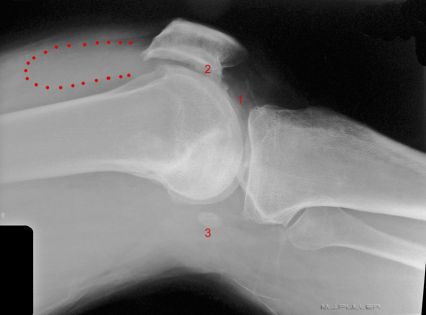
 | There is a moderately large knee joint effusion. This is seen as an increase in the size and density of the suprapatellar pouch of the knee joint (dotted red line). In addition, there is a suggestion of additional irregular fluid density seen in Hoffa’s Triangle (1). Increased density within Hoffa’s triangle/fatpad is associated with knee joint effusion. Hoffa’s fat pad is the triangular infrapatellar portion of the joint and should be an area of relative radiolucency on the lateral view (i.e. fat density rather than fluid density)
The distention of the suprapatellar pouch by the knee effusion is anteriorly displacing the quadriceps femoris muscle/suprapatellar tendon. This is causing some anterior displacement and angulation of the patella.
The irregularity of the joint surface of the patella is associated with degenerative changes(2).
There is a fabella(3). There appears to be some clothing artifact. |
Conclusion There is Pelligrini-Stieda Disease. There is a moderately large knee effusion. There is evidence of osteoarthritis. It is likely that this patient has been an active sportsman in the past. The cause of the knee effusion is unknown.
Discussion Recognition of Pellegrini-Stieda disease is a useful pattern recognition skill for radiographers who work in the Emergency Department. It is often a source of confusion for the referring doctor who may request unnecessary comparison views. Equally, recognition of a knee joint effusion either from a distended suprapatellar pouch or from fluid density in Hoffa's Triangle can be useful. Once you are able to recognise a knee effusion confidently, you will be able to recognise the absence of a knee effusion and this can be helpful in cases where there is equivocal evidence of bony injury.
....back to the applied radiography home page here


