Orthopaedic Internal Fixation Failure

From wikiRadiography
Jump to navigation Jump to search
Introduction

This page demonstrates examples of failure of internal fixation of fractures for a variety of reasons. There are no particular radiographic techniques for these types of cases other than the requirement to be mindful that patients with internally reduced and plated/pinned fractures can suffer failure of the surgical procedure.


Case 1

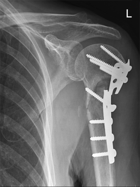
AP shoulderThe 74 year old male presented to the Fracture Clinic for routine follow-up assessment of a fractured left neck of humerus. The fractured neck of humerus had previously been surgically treated with a plate and screws.

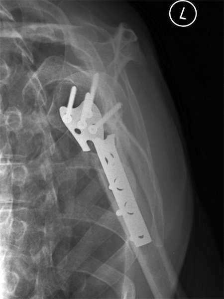
There is minimal evidence of callus formation. There is a fracture of the surgical plate.
lateral scapulaThe lateral position is over-rotated. The image appears to be under-exposed. The surgical plate is fractured.


Case 2
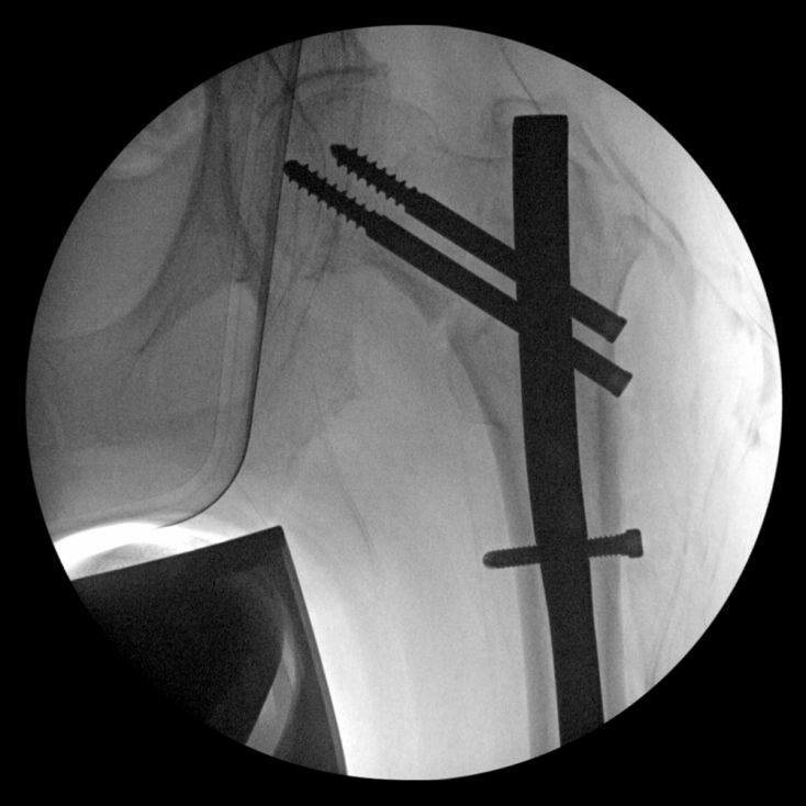
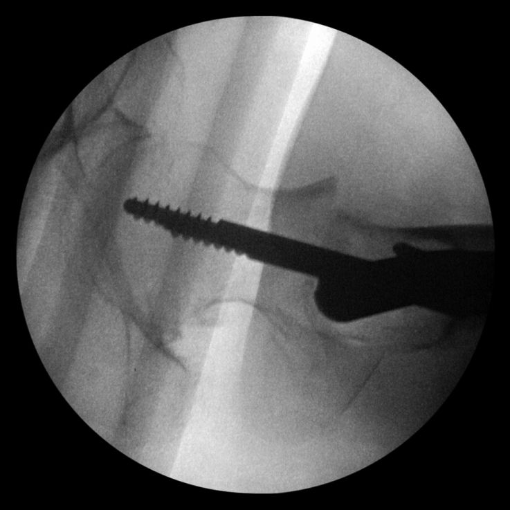
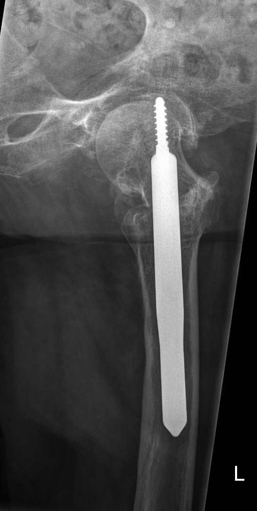
II NOF ORIFThis 93 year old lady underwent ORIF for a left neck of femur fracture. The fracture was surically treated with a cephalomedullary nail.

This is the AP image from the image intensifier as saved during the operation.
II NOF ORIFLateral of same
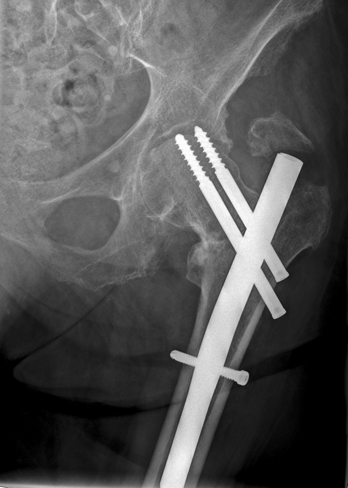
AP hipThe patient represented to hospital with unexplained left hip pain 12 months after the operation. The patient was examined and left hip radiography was requested.

The two screws have migrated superiorly with both screws encroaching on the joint.
lateral hipA rolled lateral hip technique was employed. The rolled hip projection image also demonstrates at least one of the screws in the hip joint.

Comment

This not so much a surgical hardware failure as a bone failure. Elderly female patients are particularly susceptible to bone softening associated with osteoporosis. It is the patient's weak bone that has caused this failure.