Orthopaedic Hardware
Most radiographers can recognise "orthopaedic hardware" on an X-ray image but often dont know the entire range or classifications of the hardware. This article aims to give insight into the indications and classifications of the hardware available.
Indications for Fixation
Most orthopaedic surgeons prefer to treat a fracture using a closed technique. However, sometimes this is not possible and operative fixation is necessary. The indications for operative fixation are:
- Closed methods have failed
- Closed methods will probably fail
- Displaced intraarticular fractures
- Pathological fracture
- Associated neurovascular injury
- Polytrauma
- When it will minimise confinement to bed
- When it will substantially reduce cost of treatment
Classification of Orthopedic Fixation Devices
There is a huge range of makes and models of fixation hardware however a a general classification of orthopedic fixation hardware is
- Internal Fixation Devices
- Screws
- Plates
- Wires and pins
- Intramedullary rods and nails
- Spinal fixation devices
- External Fixation Devices
- Fracture fixation
- radius
- tibia
- pelvis
- Bone lengthening
- Ilizarov device
Screws
One of the basic ideas of orthopaedic fixation is that bone heals better if the fracture fragments are pressed firmly together. Many orthopaedic devices are designed to do just that, as well as the function of stabilizing the fracture in anatomic alignment. Fracture compression increases the contact area across the fracture and increases stability of the fracture. It also decreases the fracture gap and decreases stress on the orthopedic implant. This compression can be static, where the compression is produced by the fixation device alone, or dynamic, where body weight or muscle forces are used to produce additional compression.
Screws may be used by themselves to provide fixation or in conjunction with other devices. Any screw that is used to achieve interfragmental compression is termed a lag screw. Such screws do not protect fractures from bending, rotation or axial loading forces, and other devices should be used to provide these functions.
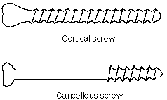
| Cortical and cancellous screws The two most common types of screws are cortical and cancellous screws. Cortical screws tend to have fine threads all along their shaft, and are designed to anchor in cortical bone. Cancellous screws tend to have coarser threads, and usually have a smooth, unthreaded portion, which allows it to act as a lag screw. These coarser threads are designed to anchor in the softer medullary bone. |
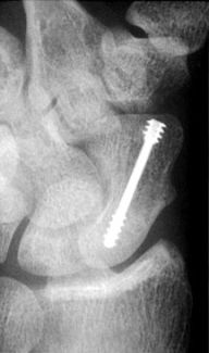
| Herbert Screw The Herbert screw is designed for use in fractures of small articular bones such as the carpals. It is cannulated and threaded at both ends. These threads run in the same direction, but the proximal portion has a wider pitch to its thread. Thus, when the proximal threads engage in the bone, they tend to move through the bone faster than the threads at the distal end, causing the two ends of the bone to compress together. This screw is used where a standard screw would impinge on adjacent tissues, such as in the treatment of scaphoid fractures. |
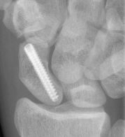
| Acutrak Screw The Acutrak screw is similar to the Herbert screw. It is also headless, which allows it to be implanted below the surface of the bone. It uses the same concept of variable thread pitch as the Herbert screw, but unlike the Herbert screw, is fully threaded. Which may improve internal holding power, as well as allow a fracture or osteotomy site to lie anywhere along the length of the screw. |
| Interference screw This screw is sometimes used in the repair of the anterior cruciate ligament (ACL). In this type of repair, the surgeon employs a cadaveric allograft ligament which has a block of bone still attached at both ends. A tunnel is drilled through the distal femur and the proximal tibia, and these bony blocks are placed within the tunnels. The interference screws are placed alongside the bone blocks so that they tightly wedge them into the side of the tunnel and prevent them from moving. |
Washers
Washers are generally used in two situations. They are used to distribute the stresses under a screw head so as to prevent thin cortical bone from splitting. Serrated washers are used to affix avulsed ligaments, small avulsion fractures or comminuted fractures to the remainder of the bone.Plates
Femoral fractures
- dynamic hip screws (DHS)Knowles pinspercutaneous pin
Wires
- Kirschner wire
- tension band wiring - patella
Rods and Nails
- Rush rod
- Ender nail
- interlocking nail
Spinal Fixation Devices
- Harrington distraction rod and compression rod
- Edwards rods
External Fixation
Antibiotic Impregnated Cement
| Antibiotic Impregnated Beads Used to deliver antibiotics by local diffusion into tissues. http://www5.aaos.org/oko/description.cfm?topic=TRA017 http://www.ajronline.org/cgi/content/full/189/2/446/FIG2 | 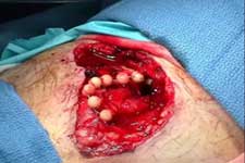 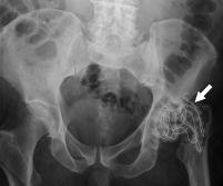 |
....Go Back to the General Radiography Homepage
