Neonatal Chest Pathology
- Hyaline Membrane Disease
- Bronchopulmonary Dysplasia
- Transient Tachypnea of the Newborn
- Meconium Aspiration Syndrome
- Neonatal Pneumonia
Hyaline Membrane Disease (Respiratory Distress Syndrome, RDS)
• Originates from insufficient surfactant production
• Inadequate surfactant production leads to high air-water (blood) surface
tensions which favour alveolar collapse and gas exchange failure in the lung
•Instability of terminal airspaces due to elevated surface forces at liquid-gas interfaces•Stable alveolar volume depends on a balance between:1)surface tension at the liquid-gas interface, and
2) recoil of tissue elasticity
• Collapsed alveoli contain fluid with a high protein content, hyaline
membranes, and lamellar bodies derived from the surfactant layer of lung[[1]]
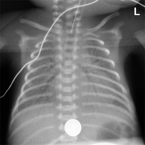
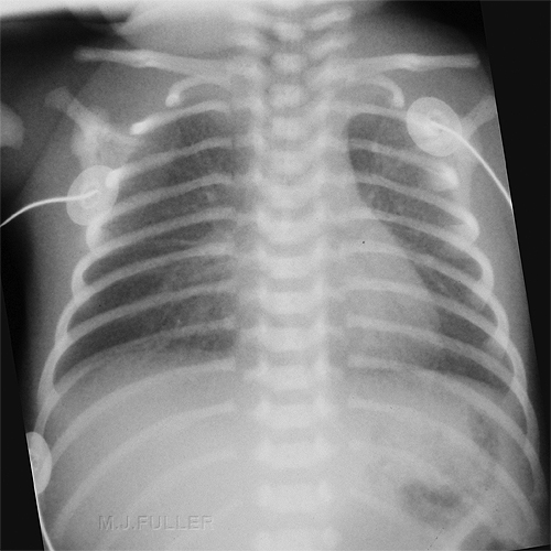
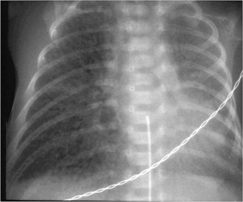
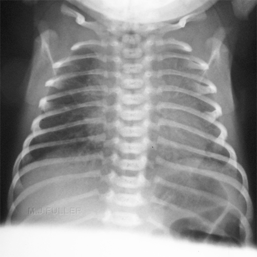
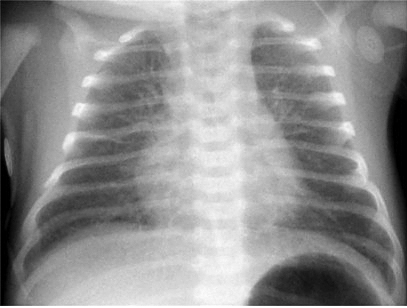
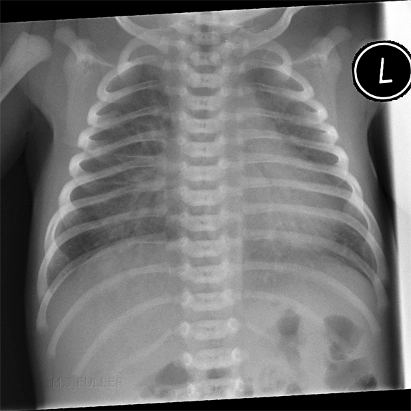
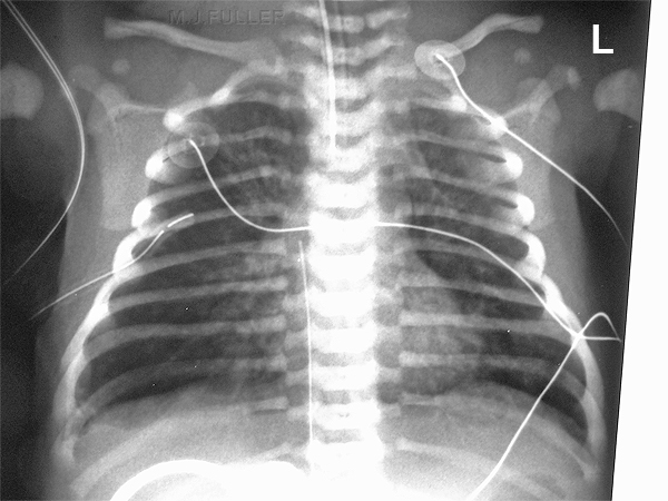
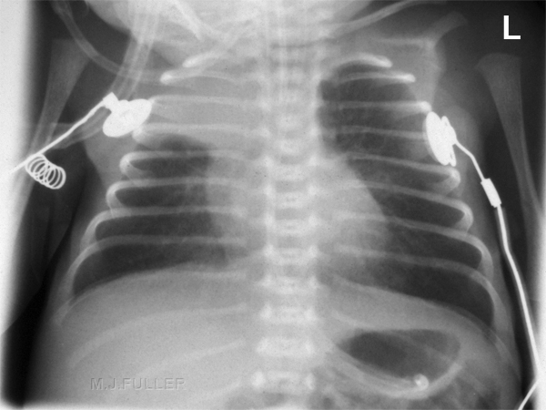
- Typically, diffuse “ground-glass” or finely granular appearance
- Bilateral and symmetrical distribution
- Air bronchograms are common
- Especially extending peripherally
- Hypoaeration in non-ventilated lungs
- Hyperinflation excludes HMD
- “Granularity” is the interplay of
- Air-distended bronchioles & ducts
- Background of atelectasis of alveoli
- May change from film-to-film if there is
- Expiration (air disappears)
- Better aeration (small bubble formation)
<a class="external" href="http://www.learningradiology.com/archives2008/COW+292-TTN/ttnccorrect.html" rel="nofollow" target="_blank">http://www.learningradiology.com/archives2008/COW%20292-TTN/ttnccorrect.htm</a>•Surfactant consists of phospholipids and protein. It begins to be produced at about 24-28 weeks of pregnancy. Surfactant is found in amniotic fluid between 28 and 32 weeks. By about 35 weeks gestation, most babies have adequate amounts of surfactant.•Surfactant is normally released into the lung tissues where it helps lower surface tension in the airways. This helps keep the lung alveoli open. When there is not enough surfactant, the alveoli collapse. As the alveoli collapse, damaged cells collect in the airways, creating a “hyaline membrane.”
chest x-rays of lungs - often show a unique "ground glass" appearance called a reticulogranular pattern.
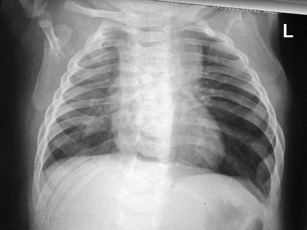
Moderately severe HMD/RDS Typical appearance of HMD/RDS
Bronchopulmonary Dysplasia
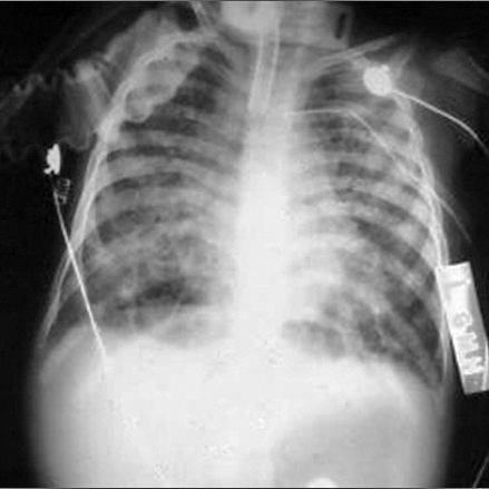
- May be impossible to distinguish early stages of BPD from later stages of HMD
- Coarse, irregular, rope-like, linear densities
- Represents atelectasis or fibrosis
- Lucent, cyst-like foci
- Hyperexpanded areas of air-trapping
- Hyperaeration of the lungs
- Conglomerate disease in BPD
- Shifting atelectasis
- Episodes of aspiration or pulmonary edema
- Superimposed pneumonia
- Changes of BPD will revert to normal on the chest radiograph in most patients after the age of two
<a class="external" href="http://www.learningradiology.com/archives2008/COW+292-TTN/ttnccorrect.html" rel="nofollow" target="_blank">http://www.learningradiology.com/archives2008/COW%20292-TTN/ttnccorrect.html</a>
Pulmonary Interstitial Emphysema (PIE)
Source: Roderick MacPherson M.D. Professor Emeritus Radiology, Medical University of South Carolina
Transient Tachypnea of the Newborn (Also known as RDS type 2 or wet lung syndrome)
- Hyperinflation of the lungs
- Fluid in the fissures
- Laminar effusions
- Fuzzy vessels
- <a class="external" href="http://www.learningradiology.com/archives2008/COW+292-TTN/ttnccorrect.html" rel="nofollow" target="_blank">http://www.learningradiology.com/archives2008/COW%20292-TTN/ttnccorrect.html</a>
•It is thought that slow absorption of the fluid in the fetal lungs causes TTN. This fluid makes taking in oxygen harder and the baby breathes faster to compensate.
•rapid breathing rate (over 60 breaths/minute)•grunting sounds with breathing•flaring of the nostrils•retractions (pulling in at the ribs with breathing)
[292-TTN/caseoftheweek292page.html|http://www.learningradiology.com/archives2008/COW%20292-TTN/caseoftheweek292page.htm]l
Meconium Aspiration Syndrome
<a class="external" href="http://www.learningradiology.com/archives2008/COW+292-TTN/ttnccorrect.html" rel="nofollow" target="_blank">http://www.learningradiology.com/archives2008/COW%20292-TTN/ttnccorrect.html</a>
- Diffuse “ropey” densities (similar to BPD)
- irregular infiltrates
- Patchy areas of atelectasis and emphysema from air-trapping
- Hyperinflation of lungs
- Spontaneous pneumothorax and pneumomediastinum
- Occurs in 25%; usually requiring no therapy
- Small pleural effusions (20%)
- No air bronchograms
- Clearing usually quick if mostly water; days-weeks if mostly meconium
- Bilateral patchy coarse infiltrates with hyperinflation
- Radiologically the typical progression is from global atelectasis in early X-rays to a widespread patchy opacification accompanied by areas of hyperinflation and/or atelectasis.
lCauses plugging of the airways with consequent atelectasislMeconium is irritating to the airways causing a chemical pneumonitis and secondary bacterial infection
<a class="external" href="http://www.learningradiology.com/archives2008/COW+292-TTN/ttnccorrect.html" rel="nofollow" target="_blank">http://www.learningradiology.com/archives2008/COW%20292-TTN/ttnccorrect.html</a>
Neonatal Pneumonia
- Perihilar streaky pattern may resemble <a class="external" href="http://www.learningradiology.com/archives2008/COW+292-TTN/ttnccorrect.html#ttn" rel="nofollow" target="_blank">TTN</a>
- Patchy airspace disease
- Diffuse, relatively homogeneous infiltrates resembling ground-glass pattern of <a class="external" href="http://www.learningradiology.com/archives2008/COW+292-TTN/ttnccorrect.html#hmd" rel="nofollow" target="_blank">HMD</a>
- Occasionally pleural effusion may occur
- Lobar consolidation from infection is unusual in a newborn
- Group B Strep looks most like HMD
- Term infant with findings of “HMD” should be considered to have pneumonia until proven otherwise
<a class="external" href="http://www.learningradiology.com/archives2008/COW+292-TTN/ttnccorrect.html" rel="nofollow" target="_blank">http://www.learningradiology.com/archives2008/COW%20292-TTN/ttnccorrect.html</a>
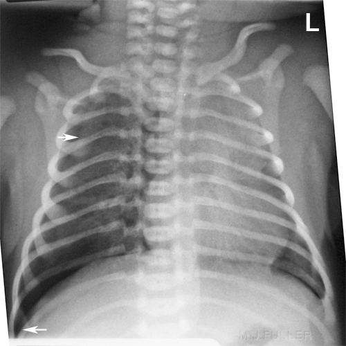
Spontaneous Pneumothorax
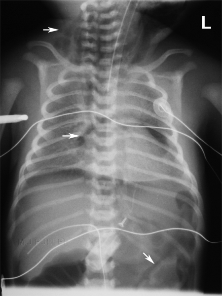
Pneumomediastinum and Pneumoperitoneum
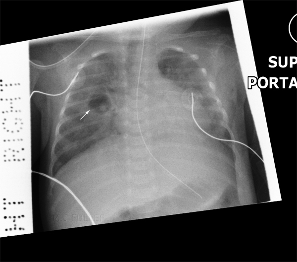
Spontaneous right pneumothorax (? tension) in newborn.
- visible lung edge (top arrow)
- deep sulcus sign
- silhouette sign right hemidiaphragm
- mediastinal shift
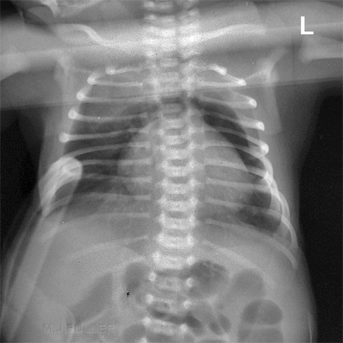
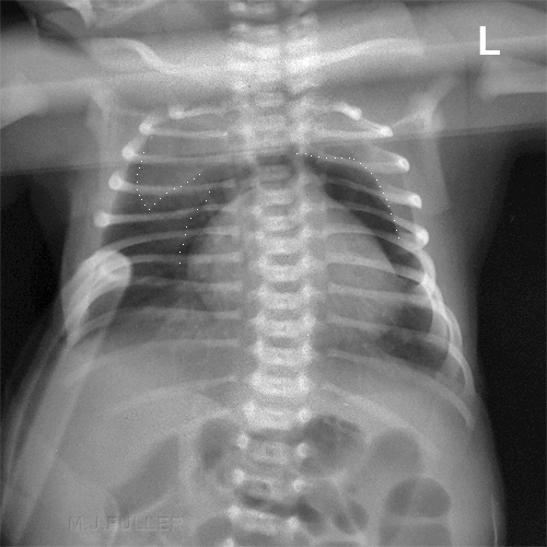
Pneumomediastinum
Right Upper Lobe Consolidation
This baby has pneumomediastinum. The dotted line in the upper right lung is the thyroid which is displaced by the pneumomediastinum. The other dotted lines outline the airfilled mediastinum
Rib anomalies
This baby has a right upper lobe consolidation with some associated collapse
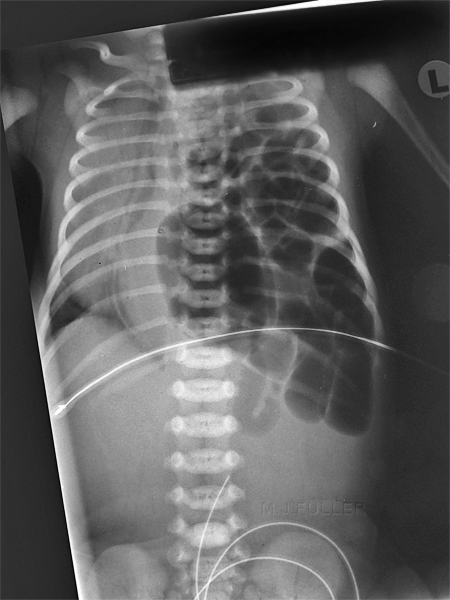
Diaphragmatic Hernia
This baby has multiple bifid/conjoined ribs.
? thoracic vertebral anomalies
Pneumatocoele
... back to the Wikiradiography home page
... back to the Applied Radiography home page
