Neonatal Chest Radiography
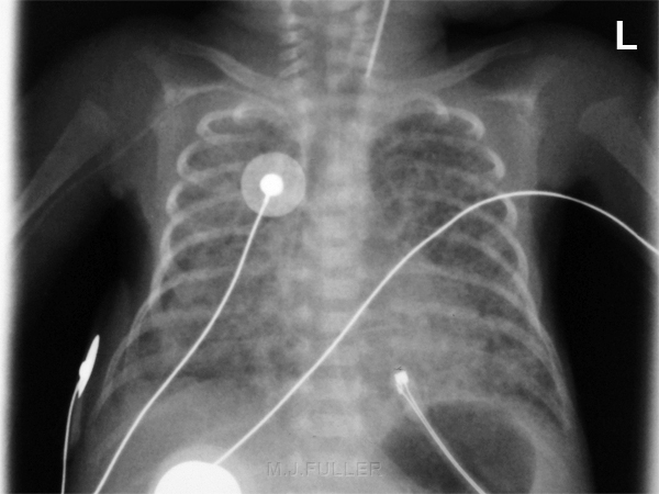
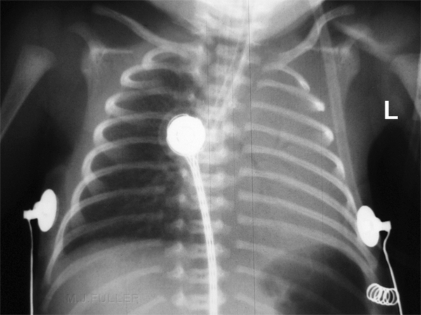
Chest radiography with the portable X-ray unit is the most commonly requested X-ray examination in the neonatal unit. Achieving high quality images can be deceptively difficult. This page examines the issues and techniques peculiar to radiography in the neonatal unit.
Abbreviations and Terminology
UAC: Umbilical artery catheter
UVC: Umbilical vein catheter
ETT: Endotracheal tube
NGT: Nasogastric tube
TPT: Transpyloric tube
VP Shunt: Ventriculo-peritoneal shunt
CVC: Central Venous Catheter
VLBW: Very Low Birthweight
NEC: Necrotising Enterocolitis
Ground Rules
You learn quickly that radiography in the neonatal unit is different. The patients are very small, vulnerable and sometimes very ill. The following are some very simple dot point tips:
- wash your hands before entering the unit and on exiting. Also, wash your hands between babies (important)
- ensure that the baby's name matches the one on the request form (careful you don't get confused with twins)
- check the clinical information on the request form- this can influence what you include on the image
- don't run the mobile machine into the cot/incubator (surprisingly easy to do)
- ask the nurses for help- they know the babies, know their illnesses, and are skilled in handling them
- be careful to avoid patient rotation, particularly with chest radiography
- use a side marker (see Baker Cone)- left and right are not always clear from the anatomy, particularly in chest radiography
- check departmental protocol re removal of ECG leads
- think about radiation protection- horizontal ray technique considerations, gonad protection, long bone protection, the nurses fingers
- you are not the boss- if the nurse says the baby can't be moved..the baby can't be moved.
- use a cassette tray if there is one built into the cot/isolette
The Patients
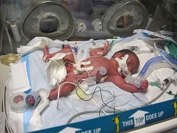
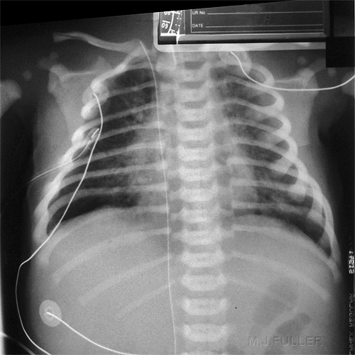
The neonatal unit patients are small- this doesn't make the job easier...in many respects it makes the job harder. The multitude of attachments is also a challenge. The attachments seem to dominate the baby.
Radiation Protection
Operator Irradiation
Operator's Hands I
Operator's Hands II
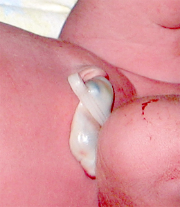
The artifact in the RLQ is the umbilicus and umbilical cord clamp
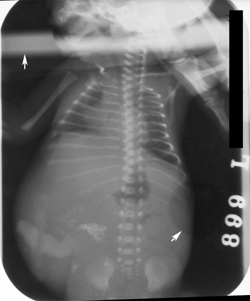
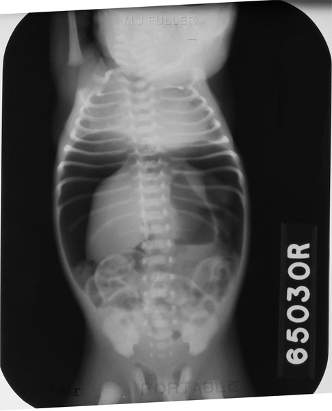
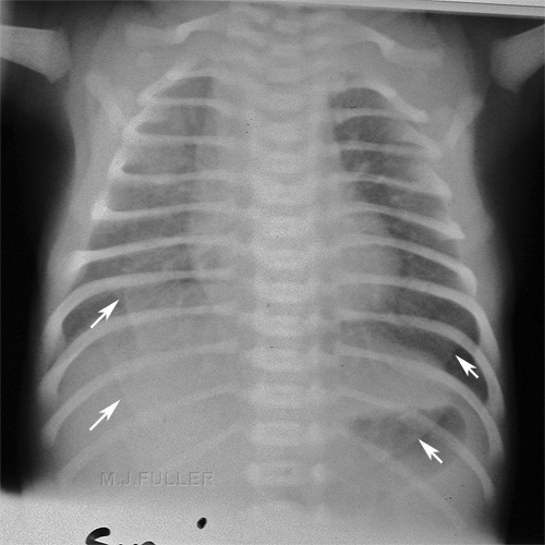
<a class="external" href="http://pregnancy.about.com/od/newbornbabies/ss/cordcare_2.htm" rel="nofollow" target="_blank">http://pregnancy.about.com/od/newbornbabies/ss/cordcare_2.htm</a>This image is circa 1966 with manual processing. Note the characteristic rounded corners of the film.
There are several valid criticisms/observations that can be levelled at this radiograph
- there is only one cone mark visible
- the operator's fingers are in the image
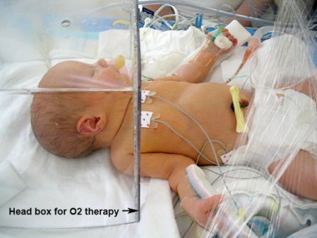
- the top artifact (white arrow) is probably a perspex head box (for O2 therapy)...see below
- there is a circular artifact that is produced by a hole in the perspex of the isolette (lower white arrow).
<a class="external" href="http://www.flickr.com/photos/71774835@N00/94691534/" rel="nofollow" target="_blank">http://www.flickr.com/photos/71774835@N00/94691534/</a>
Coning I
Coning II
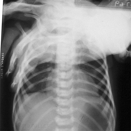
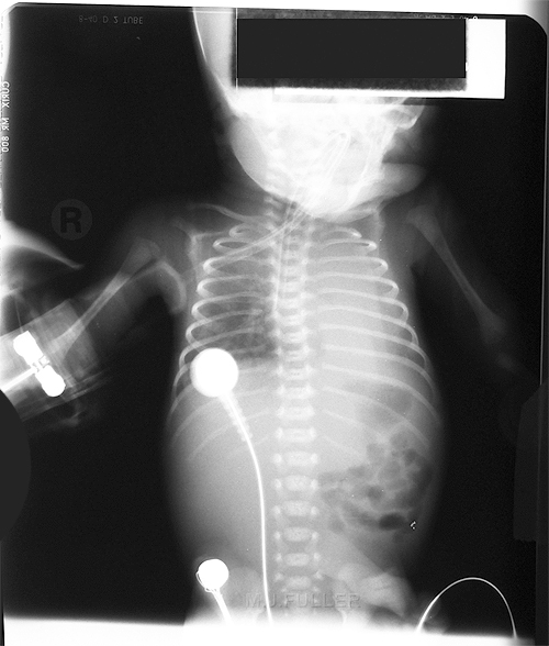
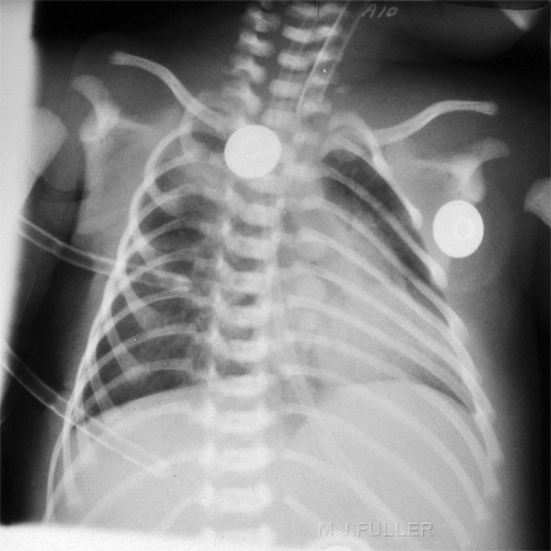
This image is circa 1970s. I suspect it is a chest X-ray and the coning is awful.
Note also
- endotracheal tube down right main bronchus
- atelectasis left lung
- skin fold over left lung
Coning III
This image is more contemporary. Baby's chests are often vaguely triangle shaped. Using Baker Cones allows the radiographer to shape the beam to the baby's chest shape. This coning may be too tight for some centres that require more of the baby's upper airway to be included in the image.
Radiographic Technique
Side Markers
Side markers in the neonatal unit are important. The difficulty is that your side marker can appear to be almost as big as the baby's chest. There are two solutions that I am aware of
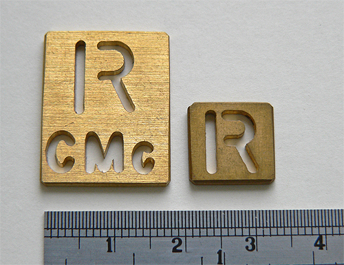
Baker ConesDedicated Neonatal MarkersBaker cones are pieces of lead rubber that have the side marker punched into them.
The marker on the left of screen is simply too large for use in the Neonatal Unit. You don't want to collimate out to include this relatively large marker. The marker on the right is the type used by the radiographers at the Adelaide Womens and Childrens Hospital. They also use these small markers with the operator's initials. Importantly, the operator's initials are engraved laterally on the marker- ie to the right on a right marker and to the left on a left marker. This ensures that if the marker is partly collimated off, it is the operator's initials that are collimated off and not the "L" or "R".
Side Markers
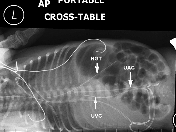
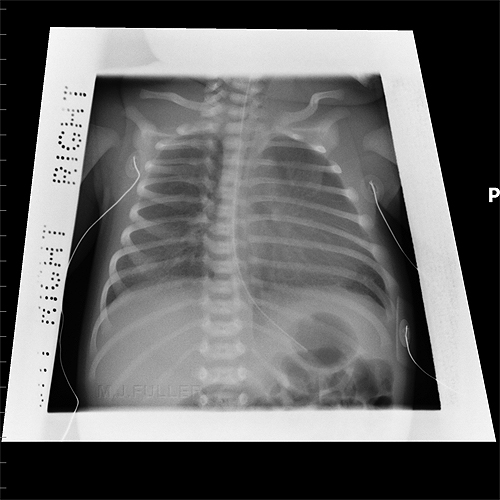
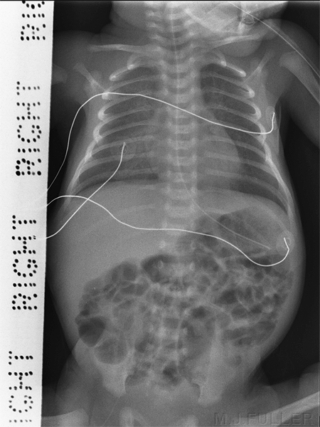
You should not rely on your ability to establish which is the correct left-right orientation of the image after it is taken. Pathology and patient rotation can make this very difficult. This patient's faintly visible left ventricle and nasogastric tube help, but what if the patient had situs inversus?
The UVC tip has tracked into the liver and there appears to be faint portal venous gas.
Air in portal venous branches can be associated with umbilical venous catheter insertion. Inconsequential transient portal venous air can be seen immediately after umbilical venous catheter insertion and should not necessarily be attributed to necrotizing enterocolitis.
<a class="external" href="http://www.ajronline.org/cgi/content/full/180/4/1147" rel="nofollow" target="_blank">Alan E. Schlesinger1, Richard M. Braverman1 and Michael A. DiPietro2 </a><a class="external" href="http://www.ajronline.org/cgi/content/full/180/4/1147" rel="nofollow" target="_blank">AJR 2003; 180:1147-1153</a>
Cassette Name Window
Exposure
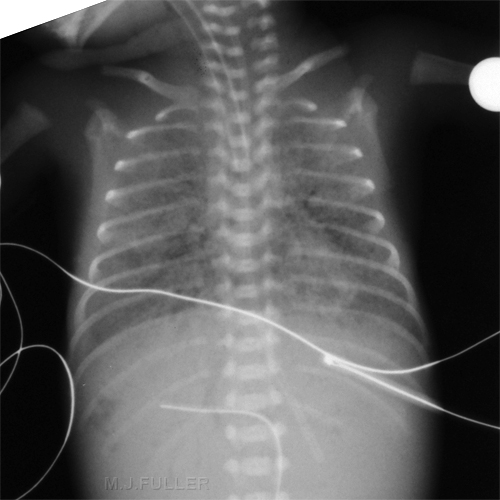
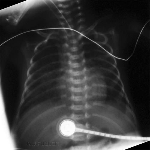
This image is overexposed. This problem tends to be masked with CR and DR systems because of their greater exposure latitude. It could be argued that the exposure used demonstrated the kinked NGT.
Skin Folds
Rotation
...back to the Wikiradiography home pageArtifacts
...baxck to the Applied Radiography home page
