Using Baker Cones in the Neonatal Intensive Care Unit
Introduction
The Neonatal Intensive Care Unit (NICU, NNU, SCBU) is an area where radiation protection is a high priority for radiographers. The Baker Cones are a customised leadrubber shield that radiographers use to restrict the primary radiation beam to the baby's chest alone. They have the added advantage of including a side marker.
Definition of Terms
Neonatal Intensive Care Unit
The neonatal Intensive Care Unit (ICU) is a department where sick and premature babies are cared for.
Lead Rubber
Lead rubber is a lead (Pb) impregnated rubber sheet that is commonly used for radiation protection applications
Light Beam Diaphragm (LBD)
The Light Beam Diaphragm is attached to the X-ray tube housing and allows the radiographer to shape the X-ray ray beam
Side Marker
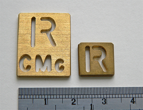
The side marker appears on the X-ray image and indicates the left or right side of the patient
History of the Baker Cone
The eponym baker cone is in recognition of the visiting radiographer from England (Jan Baker) who first introduced the idea to my department.
The Concept.
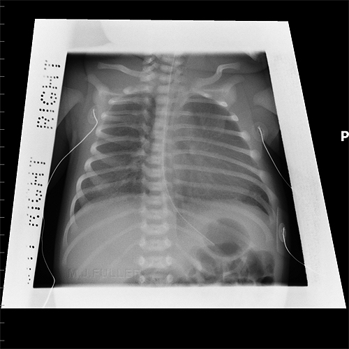
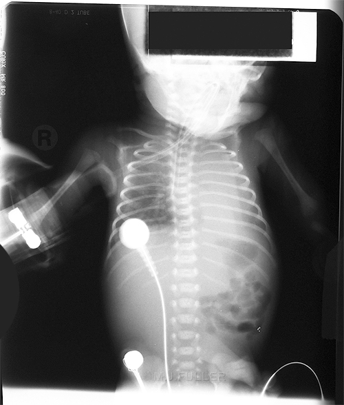
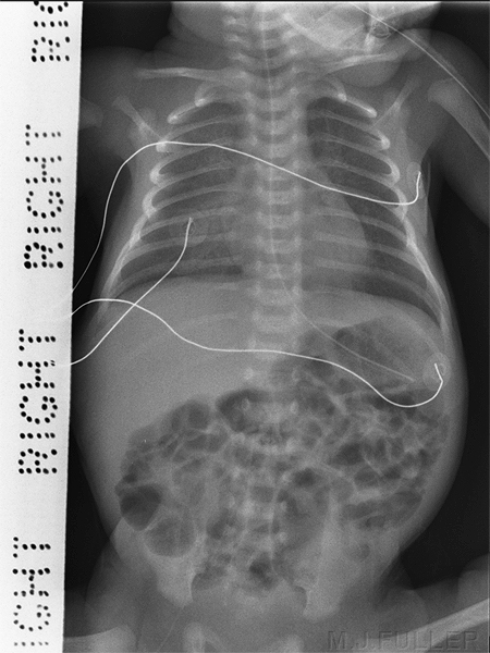
Chest radiography of premature babies can be a challenging task, particularly in non-paediatric hospitals/institutions. Two difficulties commonly arise. Firstly, it can be difficult to collimate the radiation beam such that it is restricted to the baby's chest alone- the humerii are often inadvertently irradiated. Secondly, a side marker can be difficult to place within the primary radiation field without opening the collimators or risking the side marker overlying anatomy.
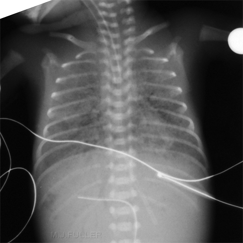
Premature babies have a chest shape that is triangular whereas the radiation field from a Light Beam Diaphragm (LBD) is rectangular. The Baker Cones allow for the primary X-ray beam to be triangular shaped and incorporate a side marker
Making a Baker Cone
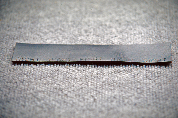
The Baker Cone is made from lead rubber with the word left or right punched along one side of the lead.
In order to obtain a professional looking result, a punch was adapted to produce consistent lettering. It was found that the holes in the lead were too small and needed reaming out by hand with a Dremel tool using a sewing needle as a reaming bit.
Technique
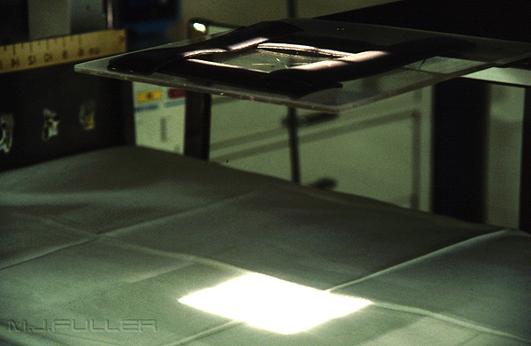
The Baker cones are positioned in the light from the LBD as shown below
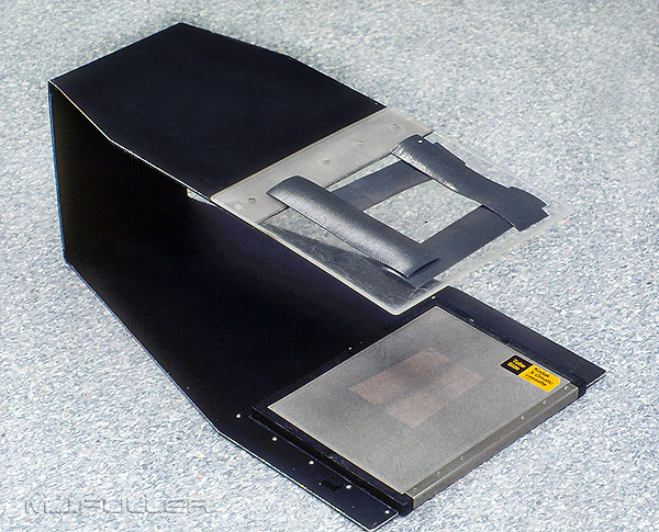
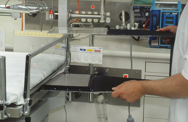
The device supporting the Baker Cones is a custom made centring device. This cassette holder is placed under the baby's mattress and has a centreing point marked on the perspex above. The device has been in use for 25 years and is universally utilised by the radiographers.
The centreing device with cassette in situ being placed under the mattress
Baker cones demonstrating the custom shaping of the LBD light
Advantages of the Baker Cone
- The baker cone serves to shape the X-ray beam into a custom shape other than the conventional rectangle. Whilst it is commonly used to prevent irradiation of the humerii, it can equally be used to create an asymmetrical X-ray field in which one of the humerii is intentionally imaged. This is sometimes undertaken for longline placement when the line enters through the cubital fossa.
- The Baker cones can be made from suitable scrap lead rubber
Disadvantages of the Baker Cone
- As with all side marker placement, care must be taken with the placement of the baker cone which incorporates the side marker perforations
- Care should be taken not to drop the Baker cones on the baby. This has not occurred to date but is clearly possible.
- The Baker cones cannot be used in open cots or incubators without the use of the pictured cassette holder.
- Baker cones may not be suitable for use on babies who are moving continuously
- The Baker Cones are made of lead rubber which is not designed to absorb primary X-ray beam. At higher kVps the cones will become less effective.
Images
The Baker Cone is seen along the baby's right chest wall. The other 3 sides use plain lead rubber
Operator's Hands
Coning I
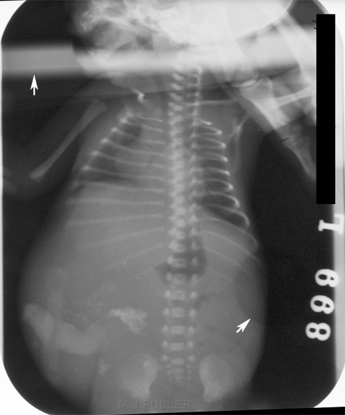
This image is circa 1961. Once again it is taken using film/screen technology and manual processing. Note that
there are no cone marks (leaves you wondering how wide the cones were).
Coning II
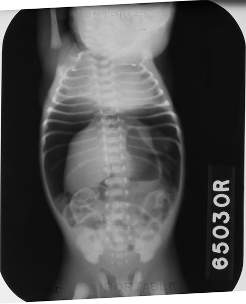
This image is circa 1970s. I suspect it is a chest X-ray and the coning is awful.
Side Markers
Side Markers
....back to the applied radiography home page here
