Modified Trauma Lateral Hip Radiography
Introduction
The Trauma HipLateral hip radiography could be usefully divided into techniques for demonstration of acute/trauma pathology and techniques for chronic pathology. This page describes three alternative techniques to the lateral horizontal ray trauma hip technique (Danelius-Miller method).
The neck of femur is a common fracture site, particularly in the elderly. Irrespective of the technique employed, an unobstructed view of the femoral neck should be achieved in the trauma lateral view of the hip. The lateral horizontal ray hip technique is commonly employed by radiographers in the trauma setting. There are a number of alternative lateral hip techniques that can be usefully employed.
Modified Rolled Lateral Hip I (Unilateral Cleaves or Frogleg Position)
Raised Knee Lateral Hip Image
Raised Knee Lateral Hip Positioning
This technique demonstrates the neck of femur clearly. This is not a true lateral view (but I'm sure you get the idea). This may be a frequent limitation of this view in patients who are in pain and unwilling/unable to externally rotate their leg any further into a true lateral position. One of the advantages of this position is that it utilizes the table bucky and benefits from the associated image quality.
Note mattress artifact.This position is similar to that employed with a rolled non-trauma lateral hip. The only difference is that the patient's knee on the affected side is raised off the table and rested on a positioning sponge. The suggested angle between the femur and the tabletop is approximately 30-35 degrees. If you increase this angle (up to 45 degrees), you will achieve even greater clearance of the trochanter off the femoral neck, however, you will also increase the elongation/distortion of the femoral neck


Raised Knee Lateral Hip
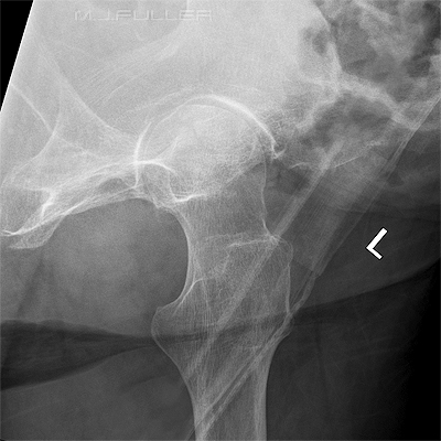
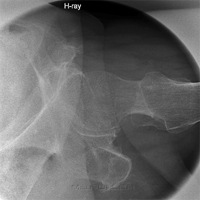
This technique demonstrates the neck of femur clearly. Compare this technique with the conventional rolled lateral hip image (right)
This is a conventional rolled lateral hip image. The greater trochanter almost completely obscures the femoral neck rendering it an unsuitable view for trauma radiography of the hip.
Raised Knee Lateral Hip Image
Conventional Lateral Horizontal Ray Trauma Hip
This technique compares favourably with a conventional lateral
horizontal ray trauma hip technique (see image on right)This is a conventional horizontal ray trauma hip image. The demonstration of the femoral neck compares favourably in this image with the "raised knee" lateral (left). Merrill's refers to this as the "Danelius-Miller method".


Modified Rolled Lateral Hip II (Friedman Method)
Modified Lateral Hip Image
Modified Lateral Hip Positioning
This technique is very similar to the previously described modified lateral hip technique. The difference is that the tube is angled instead of the anatomy (the effect is virtually identical).
The patient position is identical to a conventional rolled lateral hip. The only difference is the use of a 30-35 degree cephalic tube angle. Of course, unless you give the full 45 degrees of cephalic angle, there will be some superimposition of the greater trochanter over the neck of femur. Experience suggests that a 45 degree cephalic angle produces too much distortion of the femoral neck.


Modified Rolled Lateral Hip II (Friedman Method) at Various Tube Angulations
These images demonstrate the effect of increasing the cephalic angulation on the femoral neck. As the cephalic angle is increased the greater trochanter is increasingly projected off the femoral neck. Experience has suggested that a maximum of 35 degrees cephalic angulation provides the best compromise between projecting the greater trochanter off the femoral neck and distortion/elongation of the femoral neck.

Modified Rolled Lateral Hip III (Modified Friedman Method)
Raised Knee and Cephalic Tube Angle Lateral Hip Image
Raised Knee and Cephalic Tube Angle Lateral Hip Technique
This position is a combination of the two previous modified lateral hip techniques. The patient is positioned as shown above with a slightly raised knee (15-20 degrees) and a smaller cephalic tube angle (15-20 degrees).


Discussion
The three modified trauma hip techniques all seek to achieve the same outcome- demonstration of the neck of femur in a lateral projection unobscured by the greater trochanter. One of the quirky aspects of these techniques is that if the patient can roll their leg into the lateral position, they probably don't have a neck of femur (NOF) fracture! Patients who are referred for trauma hip radiography with a low suspicion of a fracture can often adopt this position and it is a useful positioning tool. It should also be considered that some patients with NOF fractures can present with minimal symptoms (even walking) and these patient's should not be dismissed as not having a bony injury.
One of the important advantages of these techniques is that you can use table bucky technique. Table bucky technique will usually result in a better image quality than using a horizontal ray stationary grid technique. It's also considerably more dignified for the patient than the lateral horizontal ray trauma hip technique.
...back to the Applied Radiography home page
...back to the Wikiradiography home page