IntroductionThe breathing technique is commonly employed exposure technique when undertaking lateral thoracic spine radiography. It is not clear why this technique was not adopted more widely in radiography, particularly for AP and lateral torso spine radiography. This page examines the potential benefits of a breathing exposure technique when applied to AP lumbar spine radiography.
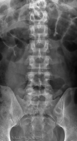
Case 1  | This 30 year old male presented to the Emergency Department following a team sports injury in which he was kneed in the lower back/right flank. The patient underwent a clinical examination and it was subsequently referred for lumbar spine radiography. Can you see any abnormal features?
There is a fracture of the right transverse process of L3. Fractures of the transverse processes of the other visualized vertebrae cannot be clearly seen, but equally cannot be excluded due to poor definition/overlap of bowel gas. The transverse processes are thin bony structures and are easily obscured by overlying bowel.
There are a few prominent air-filled (but not dilated) loops of small bowel. Some patients who are in pain air swallow, and others in severe pain develop a reflex ileus, either of which tend to obscure bony detail. |
 |
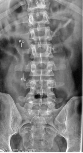
There was a suspicion that this injury was more extensive than the visualised fracture of the transverse process of L3. The AP lumbar spine image was repeated using the same kVp and mAS but using a longer exposure time of 1.6 seconds. IMAGE 2
Image(2) demonstrates the displaced fractured right transverse process of L3 clearly as well as a similar fracture at L2. I would argue that this technique should be considered in the first instance- in fact, it is equally applicable to all thoracic and lumbar spine plain film radiography (AP and lateral). The visualization of the psoas muscle is also improved.
There is a mild scoliosis concave to the right which is likely to be associated with the fractured right transverse processes.
Beware the pitfalls of breathing technique; if the patient is not compliant, you may end up with movement unsharpness of bony structures as well as soft tissues. The longer the exposure time, the greater the blurring of the soft tissue structures, but also the greater the risk of bony movement unsharpness. In short, you have to pick your patient carefully.
Was the repeated AP lumbar spine view warranted? This is a fertile area for debate. If it was known or reasonably expected that the patient would proceed to CT, the repeated view was probably not warranted. As it happens, the patient did proceed to CT and a further fractured right transverse process of L4 was identified. Fractures of the lumbar transverse processes are significantly associated with injuries to the abdominal viscera, variously reported in the literature to be between 20 and 50%. Was the repeated AP lumbar spine unnecessary in retrospect or was it influential in the decision to proceed to CT? |
Case 2 |  |
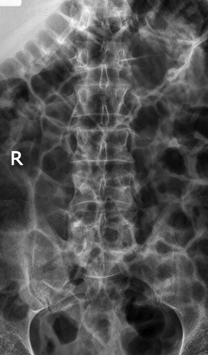
This is an AP lumbar spine image of a patient who presented to the Emergency Department following trauma. There is extensive small and large bowel gas (probably as a result of air-swallowing) which is obscuring the lumbar spine bony anatomy. The image was taken with a Philips Digital Diagnost (release 1) with default automatic exposure settings and processing algorithms.
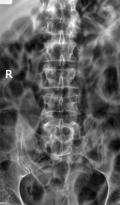
The radiographer considered that the demonstration of the bony anatomy was sufficiently poor to warrant a repeat of this view. | The image was repeated with a manual exposure technique employing the same kVp and mAS but with a fixed exposure time of 1.6 seconds. There is improved demonstration of the bony anatomy. The transverse processes of L1, for example, show improved demonstration through movement unsharpness of the overlying air-filled bowel. Some of the lumbar transverse processes remain poorly demonstrated. |
The Canadian Association of Radiologists Recommendations
The Canadian Association of Radiologists recommend breathing technique for lumbar spine radiography <a class="external" href="http://www.car.ca/uploads/standards+guidelines/lumbosacral_spine.pdf" rel="nofollow" target="_blank">http://www.car.ca/uploads/standards%20guidelines/lumbosacral_spine.pdf</a>. See also the article for the standard for thoracic spine radiography which describes why breathing technique works:
<a class="external" href="http://www.car.ca/uploads/standards+guidelines/thoracic_spine.pdf" rel="nofollow" target="_blank">http://www.car.ca/uploads/standards%20guidelines/thoracic_spine.pdf</a>
(thanks to <a href="/account/eliseleblanc" target="_self">Elise Leblanc</a> for this information)
Discussion It has been argued that you could miss an important soft tissue finding using the breathing technique. For example, a patient who presents with low back pain may in fact have renal colic and a radio-opaque stone(s) or an aortic aneurysm. It would be reasonable to ask if this situation is any different to performing a breathing technique lateral thoracic spine examination? - wouldn’t an incidental finding of bronchogenic carcinoma be equally important?
It has been argued that you should finish with one imaging modality before you proceed to the next. This argument has a certain intuitive appeal but lacks sophistication. I would argue that current practice requires radiographers to weigh up all of the relevant considerations in these situations. You should have a low threshold for consulting with the referring doctor and/or radiologist.
Good radiography is that which is the best yield/risk for that patient,
at that time, and for their likely conditions.
....back to the applied radiography home page here


