Lordotic Chest Technique
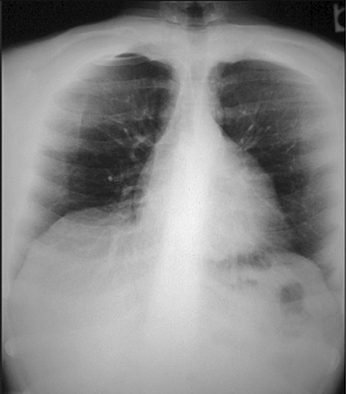
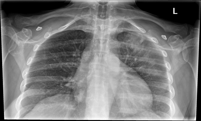
The lordotic view of the chest (first described by Felix Fleischner in 1926) is possibly underutilised because it is poorly understood (also possibly less common in an age of high resolution CT scanning). When the anatomy of interest is the lung apices, the view is sometimes referred to as an apical lordotic view- apical refers to the anatomy and lordotic refers to the patient position/technique. The lordotic technique for the apices and for the RML might only differ in their coning.The apical lordotic can be performed in the first instance if the suspected or known pathology is associated with the lung apices- eg tuberculosis. The same projection can be used to demonstrate the middle lobe of the right lung and the lingula segment of the left upper lobe.
Technique
Position
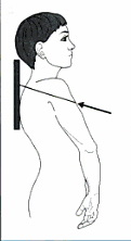
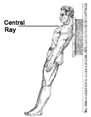
There are a variety of techniques both PA/AP and erect/supine/prone. The important factor in all approaches is that the clavicles should be projected superiorly, clear of the lungfields. My experience suggests that the exact cephalic angulation to achieve this result is debatable and variable. This may explain why the textbooks vary in their suggestions between 20 and 45 degrees of cephalic angle (or lordosis equivalent positioning with a straight tube)
This approach requires the patient to stand in an AP chest position with the central ray angled in a cephalic direction. The shoulders should be rolled forward clear of the lungfields)
adapted from Meschan, I. 1955 An Atlas of Normal Radiographic Anatomy Saunders, London
<a class="external" href="http://www.e-radiography.net/technique/chest/Chest_apical.htm" rel="nofollow" target="_blank">in http://www.e-radiography.net/technique/chest/Chest_apical.htm</a>With ambulant patients this is an alternative postioning for apical lordotic chest radiography.
source PB-322 Radiologic Technologypowerpoint presentation
X-ray Beam Coning
Lordotic Positioning for Lung Apices
Case 1
Lordotic Positioning for Right Middle Lobe and Lingula
Benjamin Felson (<a class="external" href="http://www.amazon.com/Chest-Roentgenology-Benjamin-Felson/dp/0721635911/ref=sr_1_2?ie=UTF8&s=books&qid=1252240078&sr=1-2" rel="nofollow" target="_blank">Chest Roentgenology, W.B. Saunders, 1973, p13</a>) notes "...[lordotic positioning] is extremely valuable in confirming the presence of middle lobe and lingular disease, often inconclusively demonstrated on the routine PA and lateral teleroentgenograms. In the lordotic position, the roentgen beam traverses a longer axis of the middle lobe and lingula than in the upright, producing a denser shadow when collapse is present."
<a class="external" href="http://bjr.birjournals.org/cgi/content/full/74/877/89#F6" rel="nofollow" target="_blank">
<a class="external" href="http://bjr.birjournals.org/cgi/content/full/74/877/89#F6" rel="nofollow" target="_blank"> K Ashizawa, MD, K Hayashi, MD, N Aso, MD and K Minami, MD
</a>
</a><a class="external" href="http://bjr.birjournals.org/cgi/content/full/74/877/89#F6" rel="nofollow" target="_blank">Lobar atelectasis: diagnostic pitfalls on chest radiography </a>
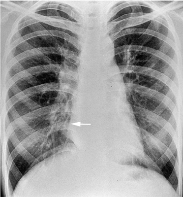
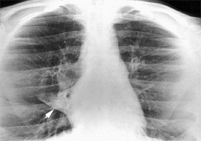
<a class="external" href="http://bjr.birjournals.org/cgi/content/full/74/877/89#F6" rel="nofollow" target="_blank">British Journal of Radiology 74 (2001),89-97 © 2001</a>The right cardiac border is not clearly seen suggesting silhouette sign associated with collapse and/or consolidation (white arrow). The appearance is a little inconclusive- the same appearance can be seen in patients with pectus excavatus
<a class="external" href="http://bjr.birjournals.org/cgi/content/full/74/877/89#F6" rel="nofollow" target="_blank"> K Ashizawa, MD, K Hayashi, MD, N Aso, MD and K Minami, MD
</a><a class="external" href="http://bjr.birjournals.org/cgi/content/full/74/877/89#F6" rel="nofollow" target="_blank">Lobar atelectasis: diagnostic pitfalls on chest radiography </a>
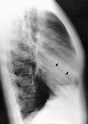
<a class="external" href="http://bjr.birjournals.org/cgi/content/full/74/877/89#F6" rel="nofollow" target="_blank">British Journal of Radiology 74 (2001),89-97 © 2001</a>the lateral image demonstrates a prominent oblique fissure (arrowed)
<a class="external" href="http://bjr.birjournals.org/cgi/content/full/74/877/89#F6" rel="nofollow" target="_blank"> K Ashizawa, MD, K Hayashi, MD, N Aso, MD and K Minami, MD
</a><a class="external" href="http://bjr.birjournals.org/cgi/content/full/74/877/89#F6" rel="nofollow" target="_blank">Lobar atelectasis: diagnostic pitfalls on chest radiography </a>
<a class="external" href="http://bjr.birjournals.org/cgi/content/full/74/877/89#F6" rel="nofollow" target="_blank">British Journal of Radiology 74 (2001),89-97 © 2001</a>The lordotic view image clearly demonstrates complete collapse of the right middle lobe (arrowed)
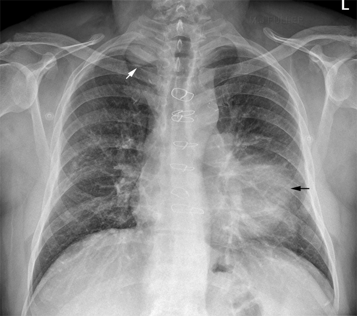
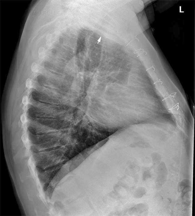
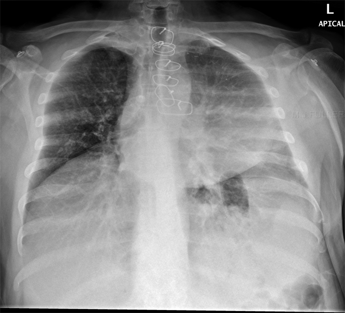
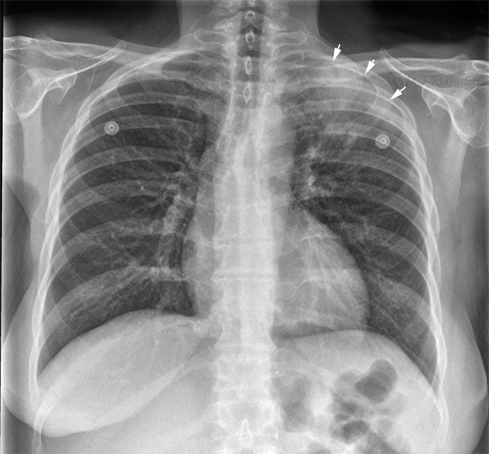
The Apical Lordotic Technique in Patients with LUL Pathology
Comment
The lordotic projections still have a place in plain film imaging of the chest. The performing of lordotic views shows thought on the part of the radiographer and will potentially be appreciated by the reporting radiologist/referring doctor.
...back to the Applied Radiography home page
