Lipohaemarthrosis

From wikiRadiography
Jump to navigation Jump to search

Introduction

Lipohaemarthrosis is a soft tissue sign that indicates the presence of an articular fracture. This page considers all aspects of imaging a lipohaemarthrosis.


Definition

A lipohaemarthrosis refers to a joint fluid accumulation that is composed of both blood and fat (lipo- fat, haem- blood, throsis- joint)

Radiography

Plain film demonstration of Lipohaemarthrosis requires a suitable exposure and a horizontal X-ray beam. Failure to employ a horizontal beam technique will either fail to demonstrate the abnormality or will suggest a joint effusion rather than a lipohaemarthrosis. The radiographer has a responsibility to check images carefully for the presence of a lipohaemarthrosis- this is particularly important when performing knee radiography in trauma patients.


Knee Effusion or Lipohaemarthrosis

Knee Effusion
Soft Tissue Signs in Orthopaedics - wikiRadiography
Lipohaemarthrosis
Soft Tissue Signs in Orthopaedics - wikiRadiography
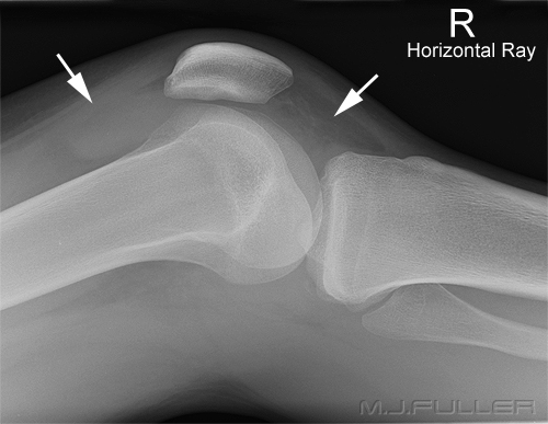
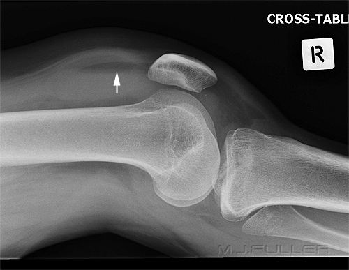
The patient fell out of a tree onto both feet. There is a knee joint effusion. This is evident from the fluid density seen in the supra-patella pouch (left arrow) and in Hoffa's fat pad (right arrow). A knee effusion indicates an intra-capsular injury (but not necessarily a fracture). A knee effusion is not particularly specific or diagnostic, but the absence of a knee effusion can be useful in cases of equivocal fracture.This patient has a lipohaemarthrosis. The fat floats on the blood forming a sharply defined blood-fat interface.


Knee Lipohaemarthrosis

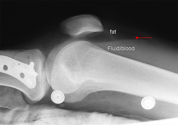
A lipohaemarthrosis refers to the presence of a blood and fat in a joint (i.e. lipo= fat, haemo = blood, throsis= pertaining to a joint). The lateral horizontal ray knee view is the one knee projection where we see this appearance. A knee lipohaemarthrosis indicates that there is a fracture that communicates with the knee joint. The blood and fat are both associated with the fracture. The fat has entered the joint from the bone medulla via the fracture. The fat 'floats' on the blood resulting in a visible interface (red arrow).

Soft Tissue Signs in Orthopaedics - wikiRadiography


It has been suggested that a patella fracture cannot cause a lipohaemarthrosis because there is insufficient fat within the patella. MR imaging of the patella (fat sat) demonstrates fat in the patella. I have seen lipohaemarthrosis associated with fractured patella on many occasions and not demonstrated any other bony injury. I am increasingly convinced that a fractured patella can result in lipohaemarthrosis of the knee which is visible on the horizontal ray lateral image.

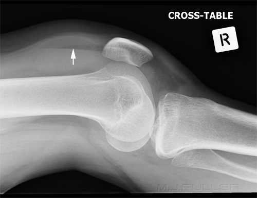
This is an erect lateral knee. A lipohaemarthrosis is demonstrated. This is a potentially useful view for subtle lipohaemarthrosis because there is no superimposed quadriceps tendon. Its usefulness is limited by the likelihood that a patient with a knee fracture will not mobilise readily.


The Snow Globe Effect
Soft Tissue Signs in Orthopaedics - wikiRadiography
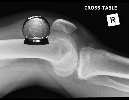
snow globe effect

adapted from <a class="external" href="http://thumbs.dreamstime.com/thumb_297/1218067161tWnq3P.jpg" rel="nofollow" target="_blank">http://thumbs.dreamstime.com/thumb_297/1218067161tWnq3P.jpg</a>
I have observed with some patients that the initial plain film image of a lipohaemarthrosis of the knee can demonstrate an indistinct fat-blood interface. One of my colleagues suggested that this could be due to the mixing of blood and fat associated with movement of the patient onto the X-ray table (or movement of the gurney/trolley/barouche/stretcher to the X-ray department). If this theory was to hold weight, the fat/fluid interface should become sharper if the image is repeated while the patient is stationary (see below)

Note also a short fat/fluid interface above the tibial tuberosity.
Soft Tissue Signs in Orthopaedics - wikiRadiographyThis is the same patient in the same position several minutes later. The fat/blood interface is now sharply defined (arrowed).
snowglobe
<a class="external" href="http://upload.wikimedia.org/wikipedia/commons/a/ab/Snow_Globe_girl.jpg" rel="nofollow" target="_blank">http://upload.wikimedia.org/wikipedia/commons/a/ab/Snow_Globe_girl.jpg</a>
A snow globe is the toy (often a souvenir) that you shake to produce an effect like falling snow.


Multi-level Lipohaemarthrosis

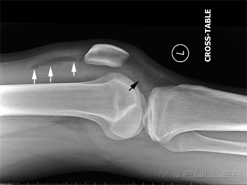
? loculated lipohaemarthrosis of the knee


There is a lipohaemarthrosis of the knee joint. There appears to be three fat-blood levels in a stepped pattern (white arrows). This may reflect some degree of normal compartmentalism of the suprapatellar space.


Lipohaemarthrosis of the Shoulder Joint
NOH fractureLipohaemarthrosis refers to a condition in which an intra-articular fracture has allowed fat to be released from bone marrow into the joint. As fat is less dense than blood, the fat 'floats' on the blood in the joint and results in an fat/blood "fluid level". This is akin to the air/fluid level seen on erect abdominal plain film images- the difference being that there are two immiscible fluids (fat and blood) rather than a gas and a fluid (air and water). With horizontal beam radiography, a fat-fluid level is detected due to differences in attenuation of these two substances.

Lipohaemarthrosis is one of the few reasonably reliable soft tissue signs of fracture. Lipohaemarthrosis is usually associated with intrarticular knee fractures but can occur (in theory) in just about any synovial joint and is occasionally seen in the shoulder joint.

Where a shoulder fracture is subtle, a lipohaemarthrosis is a useful soft tissue indicator of intra-articular fracture.

A lipohaemarthrosis will only be seen when patients are imaged with a horizontal beam (i.e. erect).

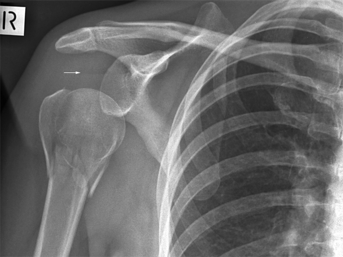
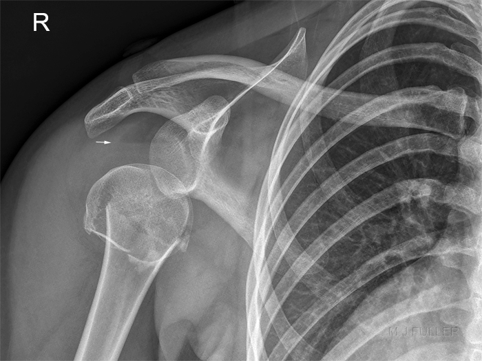
There is inferior displacement of the humeral head on the glenoid. This appearance (pseudosubluxation) is commonly seen in association with fracture and suggests joint effusion.
SHOULDER LIPOHAEMARTHROSISThis patient has a neck of humerus fracture, pseudosubluxation of the GH joint, and lipohaemarthosis.
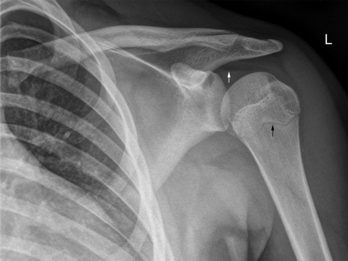
AP shoulderI suspect the white arrowed structure is a false lipohaemarthrosis. It is not a perfectly straight line and is a little more indistinct than would be expected. In addition, there is no displaced fracture demonstrated. The black arrowed structure is normal epiphysis.


Case Study 1
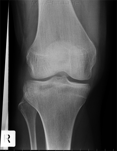
Soft Tissue Signs in Orthopaedics - wikiRadiographyThis patient presented to the Emergency Department following a sports injury the previous day. The patient sustained a blow to the knee and is now UTWB and is in great pain. The initial AP and lateral knee images are shown.


There is a suggestion of a bony defect in the tibia overlying the fibula head (not marked).
Soft Tissue Signs in Orthopaedics - wikiRadiographyThere is evidence of a lipohaemarthrosis on the lateral image (white arrow). The fat/blood interface is not demonstrated sharply. The radiographer repeated the lateral view to try and confirm the presence of a lipohaemarthrosis.
Soft Tissue Signs in Orthopaedics - wikiRadiography
The lipohaemarthrosis is now clearly seen. (the reason for the improved visualisation of the fat/blood interface on this repeat view is unclear- see snow globe effect discussion above)
Soft Tissue Signs in Orthopaedics - wikiRadiographySoft Tissue Signs in Orthopaedics - wikiRadiography
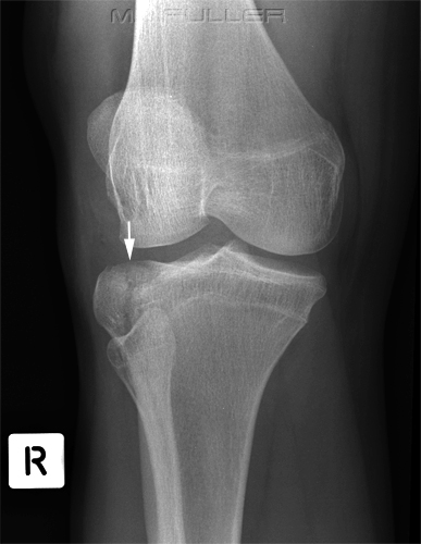
The confirmation of the lipohaemarthrosis indicates that the patient does have a fracture that communicates with the knee joint. Given that the site and nature of the fracture had not been clearly demonstrated, the radiographer proceeded to perform oblique views of the knee.

The external oblique image demonstrates a tibial plateau fracture of the lateral tibial condyle (white arrow).

Discussion
This case study demonstrates a clinical rather than a photographic approach by the radiographer. The X-ray examination was a process rather than an event. The images were taken in three stages with each stage building on the findings from the previous image(s).This is a more satisfying and effective approach than uncritically performing the routine views without further consideration of the findings.


Case Study 2
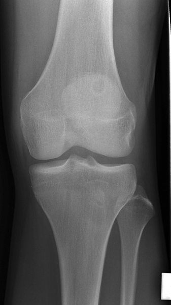
AP kneeThis 19 year old male presented to the Emergency Department after experiencing knee pain following lifting a heavy object. He was able to weightbear. He was referred for knee radiography.

There is a lytic defect with a corticated margin demonstrated in the superior aspect of the patella. This is likely to represent a normal anatomical variant.
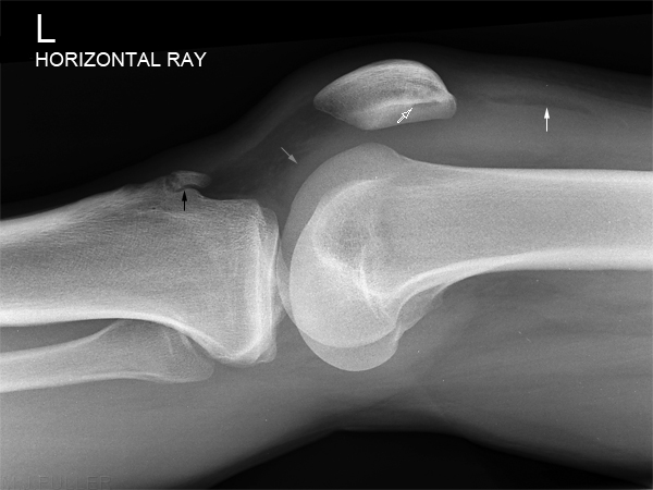
lateral kneeThere is a small lipohaemarthrosis of the knee joint (solid white arrow)

The lytic patellar defect is again demonstrated (white outline arrow)

There is evidence of a knee effusion in hoffa's fatpad. (grey arrow)

The irregularity and prominence of the tibial tuberosity is likely to represent old Osgood Schlatter's disease.
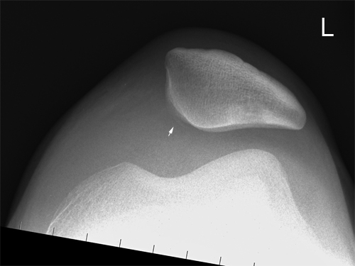
skyline kneeThe patella is demonstrated to be sited laterally. This is likey to be due to rupture of the medial patellofemoral ligament. There is also a suggestion of a bony fragment along the medial articular surface of the patella (arrowed). This may be associated with the knee joint lipohaemarthrosis.


Case 3
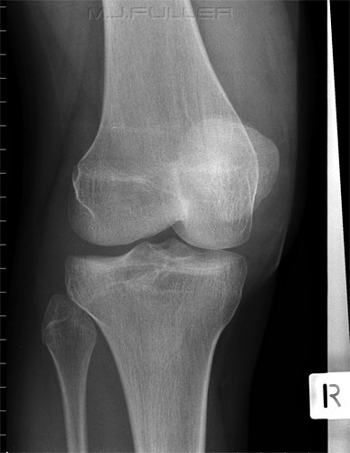
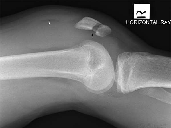
AP kneeThis 14 year old boy fell off his pushbike onto his right knee. His right knee was painful and swollen. He was referred for right knee radiography.

There appears to be a lucent line through the medial aspect of the patella.
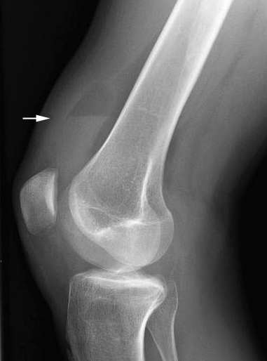
lateral kneeThe horizontal ray lateral knee view demonstrates a fractured patella and lipohaemarthrosis.


Case 4

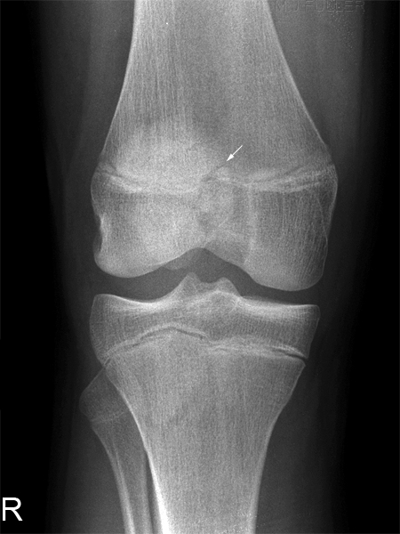
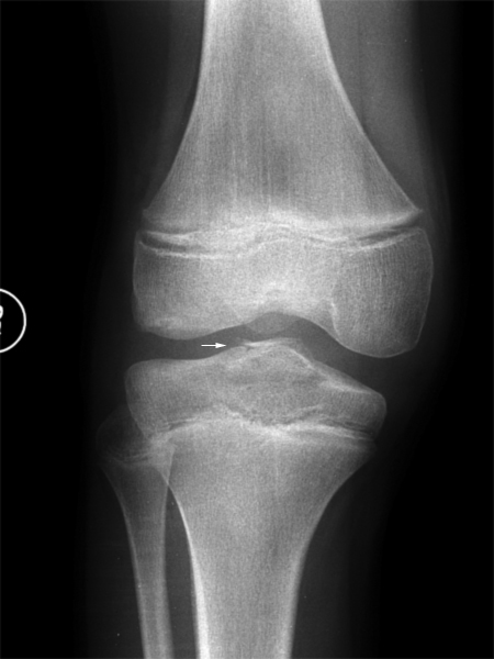
AP kneeThis 10 year old boy presented to the Emergency Department folowing a fall from his pushike. His right knee was painful and swollen. He was referred for right knee radiography.

There is irregularity of the tibial eminance (arrowed)
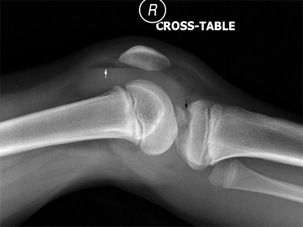
lateral kneeThe lateral view image demonstrates a lipohaemarthrosis (white arrow)

There is an avulsion fracture of the tibial eminence (black arrow)
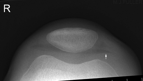
skyline kneeThe skyline image demonstrates a lipohaemarthrosis (white arrow).


Case 5

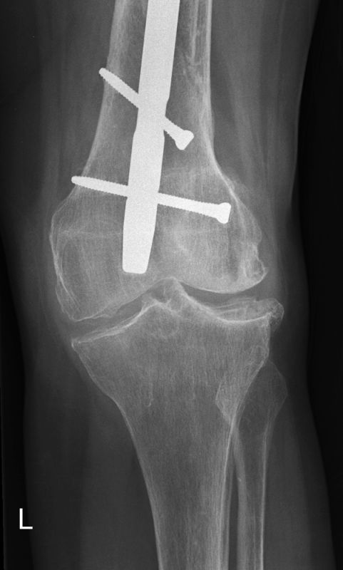
AP kneeThis 84 year old lady presented to the Emergency Department following a fall. She was examined and referred for a cocktail of imaging examinations including left knee.

The AP projection of the left knee demonstrates an intramedullary femoral nail with two screws. There is a double cortical appearance on the medial aspect of the distal femur. It is not clear if this represents an acute bony injury.

The knee joint demonstrates degenerative changes.
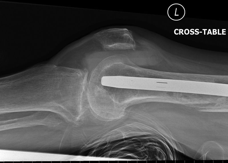
lat knee lipohaemarthrosisThe lateral projection horizontal ray image demonstrates a lipohaemarthrosis. The lipohaemarthrosis consists of 3 layers- this is of unknown aetiology. The distal femoral cortical discontinuity appears more likely to be a recent fracture in this projection, but this fracture is not associated with the knee joint lipohaemarthrosis.
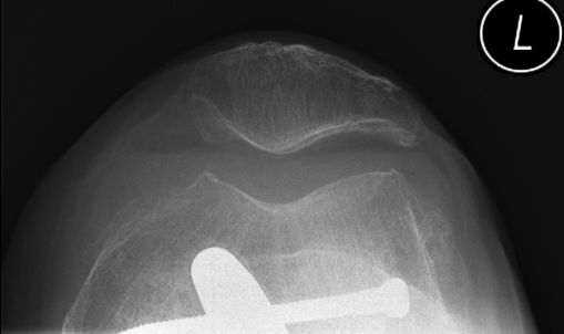
skyline patellaThe skyline projection demonstrates the lipohaemarthrosis.


References

1. Weissman, B and Sledge, C. Orthopedic Radiology. Saunders and Company, 1986

2. Hall FM. Radiographic diagnosis and accuracy in knee joint effusions.
Radiology 1975;1 15:49-54


... back to the Applied Radiography home page