Lateral Sternum from Lateral Chest- Digital Double Dipping
Introduction
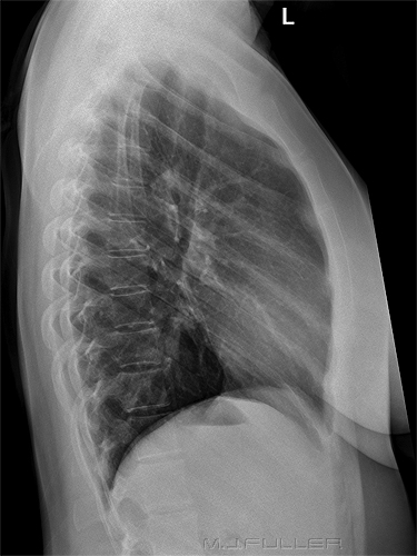
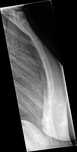
With the introduction of digital radiography, radiographers are able to potentially create 2 images from a single exposure using post-processing manipulations. One example of this digital double dipping is to create a lateral sternum image from a lateral chest image.
Is this a legitimate technique? What are its limitations?
TechniqueThere is little doubt that this double dipping can be achieved successfully and is appropriate in some circumstances. However, limitations of the technique should be appreciated.
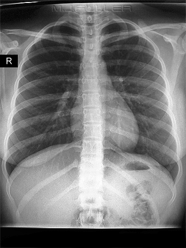
Case Study 1This patient presented to the Emergency Department following a motor vehicle accident. She was complaining of a painful sternum and was referred for radiography of the chest and sternum.
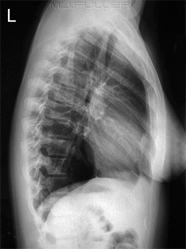
The radiographer has positioned the patient with her arms folded across the top of her head instead of the "elbows forward" position. This maximises the visualisation of the upper chest and sternum. The radiographer has used the "paravertebral gutter technique" to line up the posterior ribs which are largely superimposed. This is a technique for achieving a lateral chest position which is employed by some radiographers. The advantage of this technique is that identical lateral chest images can be produced, increasing the comparability of the images. The disadvantage of this technique is that the chest is technically not lateral. Also, the posterior costophrenic recesses can be difficult to distinguish, particularly in children. A further disadvantage is that in lining up the posterior ribs with the divergent beam, the sternum is rotated off the lateral position. If you look at the sternum in the lateral chest image above, it is not clear if the sternum is oblique, or if the patient has mild pectus excavatus, or both.
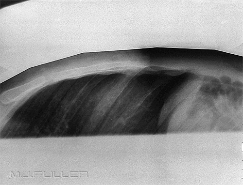
The radiographer considered the visualisation of the sternum on the lateral chest image to be inadequate and performed a dedicated lateral cross-table sternum projection using the snake to reduce scatter radiation.
Whilst this image has some shortcomings, it does demonstrate that the lateral chest image above did not demonstrate the patient's sternum in the lateral position.
Case Study 2
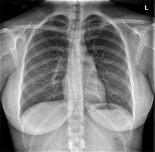
This patient presented to the Emergency Department following a motor vehicle accident. She was complaining of a painful sternum and was referred for radiography of the chest and sternum (yes.... same history as the previous case study)
The radiographer considered the visualisation of the sternum on the lateral chest image to be inadequate for the lower sternum. A dedicated erect lateral sternum was performed with a view to improving the visualisation of the lower sternum.
The dedicated lateral sternum image has achieved marginally improved visualisation of the lower sternum.
SummaryThe possibility of extracting two images from a single exposure should always be considered when using digital radiography. The limitations of the technique should be balanced against all of the relevant considerations at the time of the examination. A knowledge of the limitations of digital double dipping will help the radiographer to arrive at a well balanced decision.
....back to the applied radiography home page here
