Lateral Lumbar Spine Breathing Technique

From wikiRadiography
Jump to navigation Jump to search

Introduction

There has been a longstanding practice in radiography to perform lateral thoracic spine radiography using a long exposure time to blur out lung and other soft tissue markings. It is unclear why this technique was not seen as equally applicable to the AP projection. Equally, there are patients who present for lumbar spine radiography who have so much small bowel gas that the bony anatomy is almost completely obscured when utilising a short exposure time technique. Why not employ breathing technique for these patients?


Breathing Technique Case Study 1

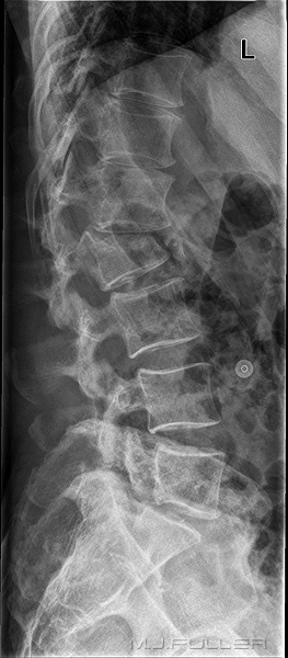
This patient has presented to the Emergency Department following a motor-vehicle accident. The patient was referred for lumbar spine radiography.

First PresentationPost Operative Imaging
Lateral Lumbar Spine Breathing Technique - wikiRadiographyLateral Lumbar Spine Breathing Technique - wikiRadiography
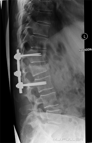
There is a crush fracture of the body of L2. The patient was referred for a surgical assessment and received pedicle screws to stabilise the fracture.

The radiographer set the kVp at 85 and the automatic exposure device determined the exposure mA and time. The exposure was made on arrested respiration. Note that there are bowel gas and diaphragmatic shadows overlying the spine.

The patient returned to the X-ray department following spinal surgery for post-operative imaging. The radiographer selected the following manual exposure factors

  • 85 kVp
  • 40 mA
  • 2.0 sec


The long exposure time has resulted in blurring of the bowel gas, ribs and diaphragm. The spinal bony anatomy remains sharp. The patient was asked to hold still but remain breathing during the exposure. This technique should not result in an increase in the mAS used- the mA is reduced to match the increased exposure time.


Breathing Technique Case Study 2

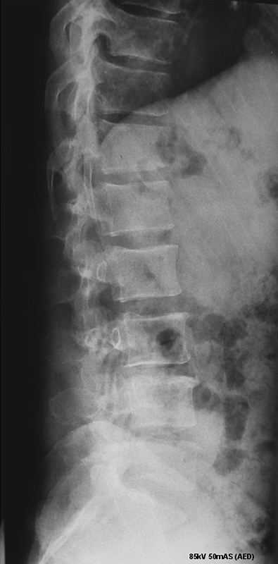
This patient has presented to the Emergency Department with back pain following a fall. The patient has a history of disseminated cancer and was considered to be at risk of a pathological fracture. The patient was referred for lumbar spine radiography with special emphasis on the L3- L5 vertebra.


Lateral Lumbar Spine Breathing Technique - wikiRadiographyLateral Lumbar Spine Breathing Technique - wikiRadiography
The lateral spine radiograph was performed using the automatic exposure chamber at 85 kVp. An acceptable exposure was achieved but the area of interest is partially obscured by bowel gas. The mAS was determined by the AEC device and was recorded as 50 mAS.

The radiographer considered that the area of interest (L3 - L5) was sufficiently obscured by bowel gas to warrant a repeat view.
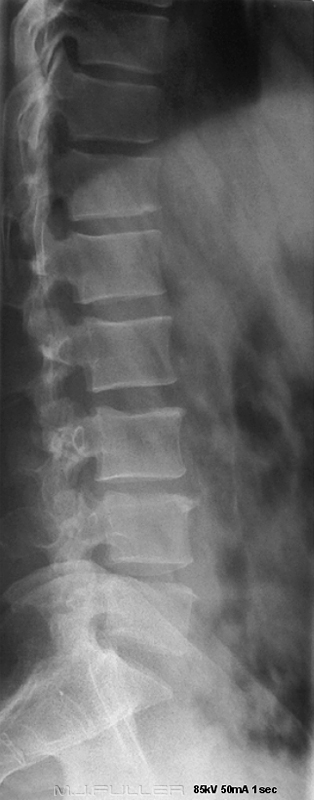
The repeat radiograph was performed using a manual exposure technique as follows

85kVp 50mA 1.0 sec

This is the same mAS as used previously but with a set exposure time of 1.0 seconds. The area of interest is now demonstrated without the detracting overlying bowel gas. This repeat image is more likely to demonstrate a pathological fracture in the area of interest that the original lateral.

There is no clearly demonstrated crush fracture or other acute pathology. There is minor osteophytic lipping and some minor wedging of the T12 vertebral body.

It is noteworthy that the spinous processes have been inadvertently collimated off.


Breathing Technique Case Study 3
This patient presented to the Emergency Department with back pain following a 2.5metre fall. The patient was complaining of hip and back pain. The patient was referred for hip and lumbar spine radiography.

AP Lumbar SpineThe radiographer considered the risk of rolling the patient too high given the possibility of a lumbar spine or pelvis fracture. The patient was slid onto the DR table and an AP breathing lumbar spine technique was employed (image left).

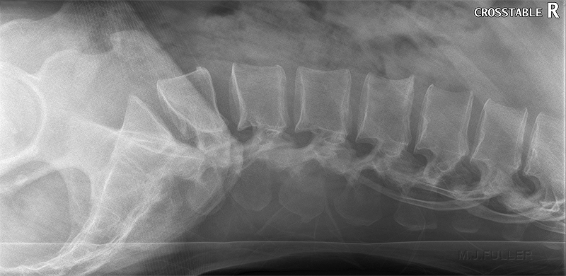
Although there was no evidence of a lumbar spine fracture on the AP view, it was considered prudent not to roll the patient for the lateral lumbar spine projection. The patient was slid back onto the barouche and a horizontal ray breathing lateral lumbar spine technique was employed using the DR vertical image receptor (image below). The lateral horizontal ray image demonstrates that this technique does not necessarily result in a sub-standard image. Importantly, this technique frequently fails if the patient is lying on a soft mattress. If you slide the patient back onto the barouche using a slide board, it is worth considering whether you can leave the slide board under the patient until the horizontal ray image has been checked.
lat lumbar spine


Breathing Technique Case Study 4
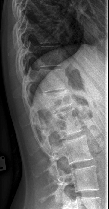
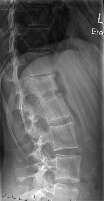
These two images are follow-up lateral lumbar spine images on a patient who sustained a crush fracture of T12
lateral lumbar spinelateral lumbar spine
This is a CR image performed using the default automatic exposure device on arrested respiration.This is the same patient imaged using a Philips DR system (reslease 2). The radiographer has selected a manual breathing exposure technique using an exposure time of 1.0 second and a fixed mA and kVp. The combination of the DR technology and the breathing exposure technique provides improved demonstration of the patient's crush fracture.

(note that the CR image is probably underexposed resulting in a low signal to noise ratio- i.e. operator error rather than technology shortcoming)


Breathing Technique Case Study 5
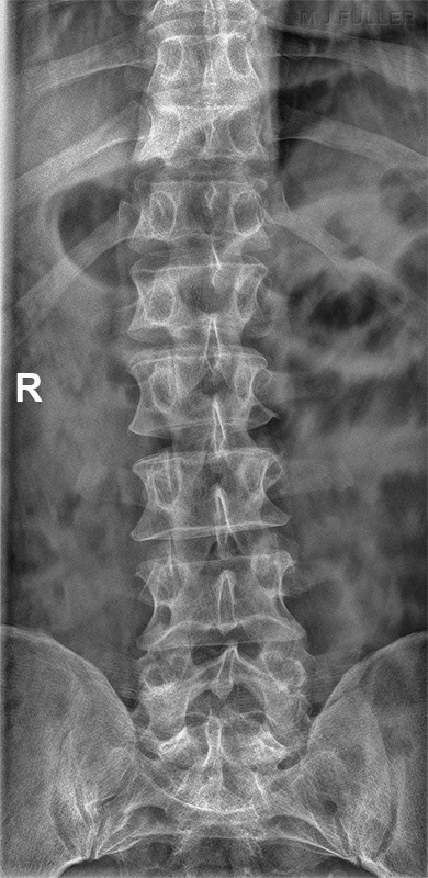
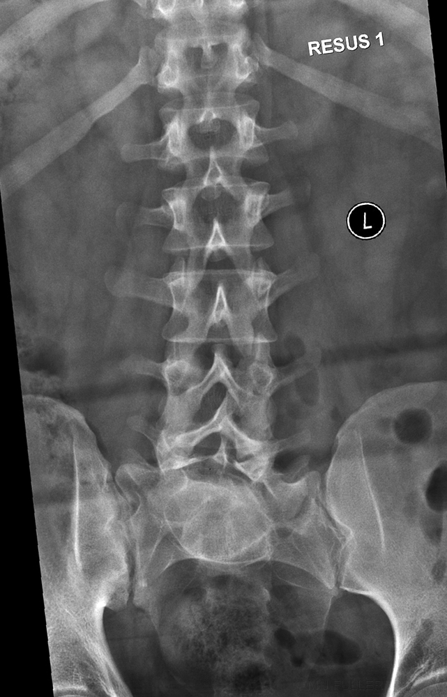
AP LUMBAR SPINEThis 31 year old male presented to the Emergency Department after falling off a ladder. A trauma series was requested including lumbar spine.

The AP projection image demonstrates the use of breathing technique. The exposure factors are unknown. The transverse processes are demonstrated particularly well.
This is a CR image performed using the default automatic exposure device on arrested respiration.


Summary

A breathing exposure technique can be employed for all torso spine radiography. It comes with an increased risk of unwanted movement unsharpness and should be used judiciously after assessing the patient's likelihood of holding still during the exposure. The longer the exposure time, the greater the blurring of the soft tissue structures and the greater the likelihood of movement unsharpness.


....back to the applied radiography home page here