Knee Trauma1
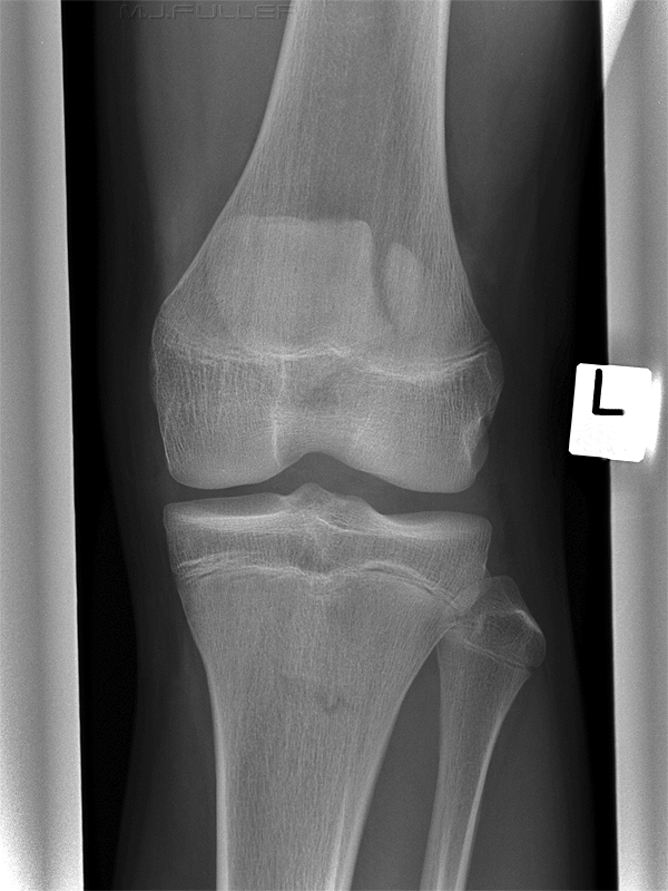
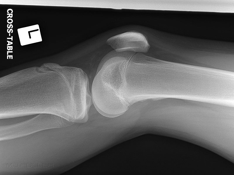
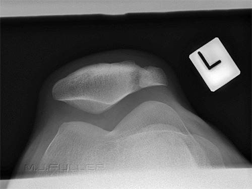
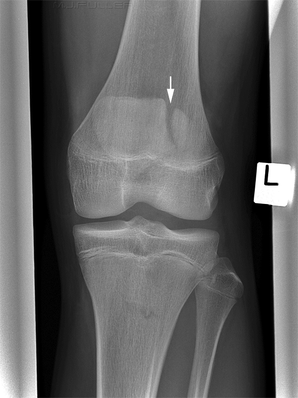
This boy presented to the Emergency Department following a fall from his pushbike. He was examined and was found to have a tender left knee and shoulder. He was referred for left knee and left shoulder radiography. The left knee images are shown below.
Images
Findings
| | There is a vertical defect in the patella (white arrow). Consideration should be given to a patella fracture or a bipartite patella (normal anatomical variant) It appears that the bony margins of the defect in the patella are smooth and the corners are radiused. This is a feature of a bipartite patella rather than a fracture. Examination of the lateral and skyline views should help to clarify the diagnosis |
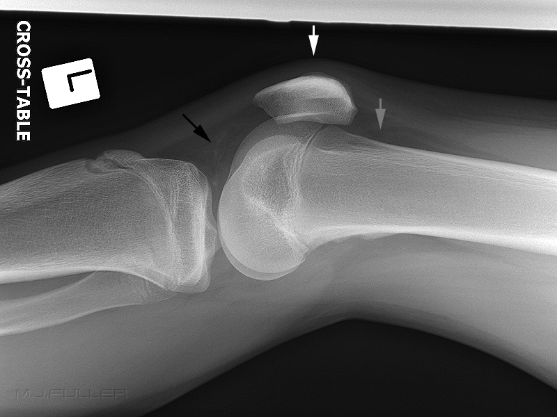 | If the patient had sustained a fractured patella there should be considerable associated soft tissue swelling. The tissues overlying the patella appear normal (white arrow) There is also likely to be a knee effusion or lipohaemarthrosis in the presence of a patellar fracture. The suprapatellar pouch appears normal (grey arrow). Hoffa's fat pad does not suggest that the patient has a significant knee joint effusion. |
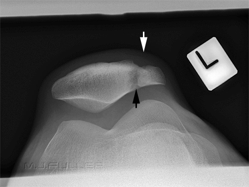 | Once again, the soft tissue contours associated with the patella appear normal (white arrow) The defect in the patella is again clearly seen. There is no evidence of sharply defined bony margins that you would expect with a fractured patella. |
Discussion
The appearances strongly suggest that this patient has a bipartite patella. If there had been coincidental overlying soft tissue swelling, the diagnosis of bipartite patella may be more difficult. However, the lack of a knee joint effusion, smooth bony contours associated with the defect and unconvincing clinical signs should not lead to a diagnosis of patellar fracture.
If the referring doctor was convinced that the appearances were consistent with a patella fracture, what would you do to clarify the diagnosis?
You may be lucky enough to find that the contra-lateral patella was similarly bipartite (less than 45% of patients will have bilateral defects). A better strategy would be to find similar appearances in Keat's " Atlas of Normal Roentgen Variants That May Simulate Disease". If you are only have one textbook in your Emergency Department, this is the one I would choose. I refer to this book on a daily basis.
...back to the Applied Radiography home page
