Introduction to the Skin
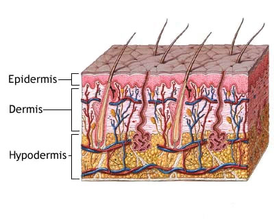
The skin is considered the largest organ of the body and has many different functions. The skin functions in thermoregulation, protection, metabolic functions and sensation. The skin is divided into three main regions, the epidermis, the dermis and the hypodermis, each providing a distinct role in the overall function of the skin. The dermis is attached to an underlying hypodermis, also called subcutaneous connective tissue, which stores adipose tissue and is recognized as the superficial fascia of gross anatomy.
Epidermis
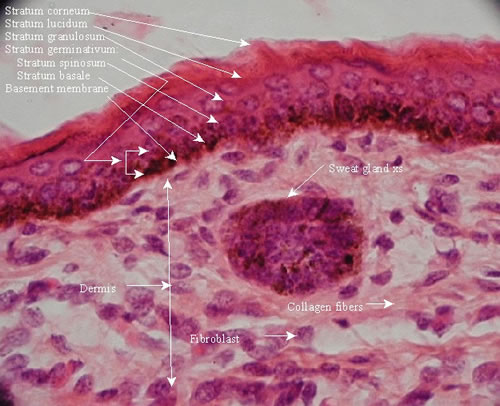
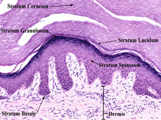
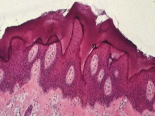
The epidermis is the most superficial outer layer of the skin and provides the first barrier of protection from the invasion of foreign substances into the body. The principal cell of the epidermis is called a keratinocyte. The epidermis is subdivided into five layers or strata, the stratum basalis(SB), the stratum spinosum(SS), the stratum granulosum(SG), the stratum lucidum (SL) and the stratum corneum(SC) in which a keratinocyte gradually migates to the surface and is sloughed off in a process called desquamation.
Stratum Spinosum
The cells that divide in the statum germinativum soon begin to accumulate many desmosomes on their outer surface which provide the characteristic “prickles” (seen on the close-up view) of the stratum spinosum (SS), which is often called the prickle-cell layer.
Stratum granulosum
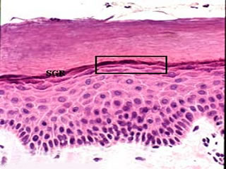
The progressive maturation of a keratinocyte is charcterized by the accumulation of keratin, called keratinization. The cells of the stratum granulosum (SG) accumlate dense basophilic keratohyalin granules (seen on the close-up view). These granules contain lipids, which along with the desmosomal connections, help to form a waterproof barrier that functions to prevent fluid loss from the body.
Stratum Lucidum
Epidermis varies in thickness throughout the body depending mainly on frictional forces and is thickest on the palms of the hands and soles of the feet. The stratum lucidum is normally only well seen in thick epidermis and represents a transition from the stratum granulosum to the stratum corneum.
Stratum corneum
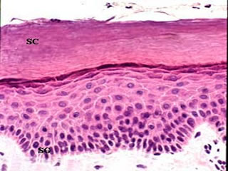
As a cell accumulates keratinohyalin granules, it is thought that rupture of lysosomal membranes release lysosomal enzymes that eventually cause cell death. The dead and dying cells filled with mature keratin form the stratum corneum (SC). The deeper cells of the stratum corneum retain their desmosomal junctions, but as they are pushed to the surface by newly forming cells of the stratum germinativum (SG), the dead cells gradually break apart and are lost, a process called desquamation.
Dermis
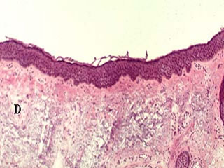
The dermis (D) assumes the important functions of thermoregulation and supports the vasular network to supply the avascular epidermis with nutrients. The dermis is typically subdivided into two zones, a papillary dermis and a reticular layer. The dermis contains mostly fibroblasts which are responsible for secreting collagen, elastin and ground substance that give the support and elasticity of the skin. Also present are immune cells that are involved in defense against foreign invaders passing through the epidermis.
Papillary dermis
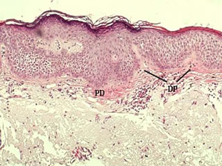
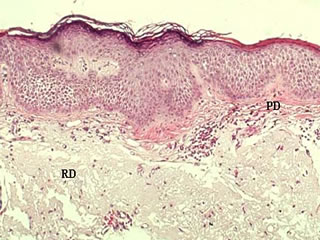
The papillary dermis (PD) contains vascular networks that have two important functions. The first being to support the avascular epidermis with vital nutrients and secondly to provide a network for thermoregulation. The vasculature is organized so that by increasing or decreasing blood flow, heat can either be conserved or dissipated. The vasculature interdigitates in areas called dermal papillae (DP). The papillary dermis also contains the free sensory nerve endings and structures called Meissner’s corpuscles in highly sensitive areas
Reticular dermis
The reticular layer of the dermis (RD) consists of dense irregular connective tissue, which differs from the papillary layer (PD), which is made up of mainly loose connective tissue. The reticular layer of the dermis is important in giving the skin its overall strength and elasticity, as well as housing other important epithelial derived structures such as glands and hair follicles.
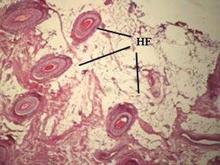
Skin Appendages
The skin contains a variety of appendages, mainly hair follicles (HF), sweat glands , and sebaceous glands, which are all embrylogically epidermal in origin.
◄.....
Go Back to Gross Anatomy Home Page 


