Imaging Subpulmonic Effusions
A pleural effusion is an abnormal condition in which fluid has accumulated in the pleural space. The plain film signs of pleural effusion are commonly seen and readily identified on plain film chest images.
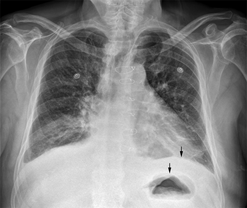
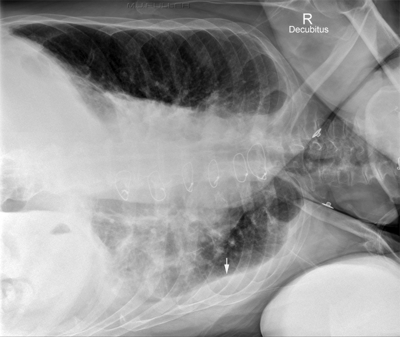
Pleural fluid can accumulate in isolation between the lung base and diaphragm. This is known as a subpulmonic effusion and presents a more difficult diagnostic challenge. When a chest X-ray is reported by a radiologist as "normal", there is potential for embarrassment when that slightly elevated-looking diaphragm turns out to be a large subpulmonic effusion. The radiographer can help prevent such embarrassment with the selective application of the decubitus chest projection on patients with suspected subpulmonic effusion.
The Pattern Recognition
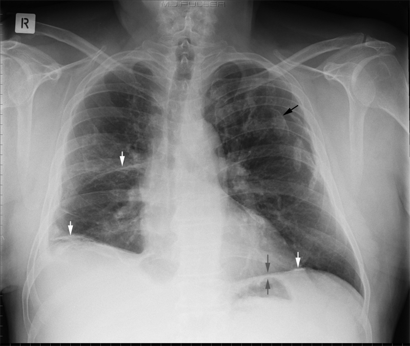
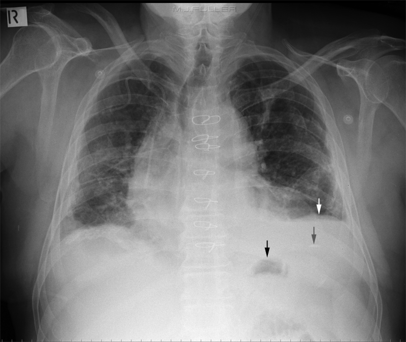
Sign Comment Abnormally large distance between fundus of stomach and lung base - erect chest only
- must have air-filled fundus of stomach
- only identifies pathology on left"abrupt termination of vascular shadows at the level of the diaphragm" (1) - may be somewhat exposure dependent (you may see nothing under the diaphragms on a slightly underexposed radiograph for example) Blunting of affected costophrenic angle (PA) - usually only seen with larger effusions A blunted posterior costophrenic sulcus may be seen on the lateral film Pseudodiaphragm can appear to "peak" more laterally Pseudodiaphragm can appear more horizontal medially than you would typically see with a normal diaphragm the medial pseudodiaphragm almost always looks abnormal Crowding of lung parenchyma on affected side
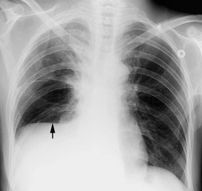
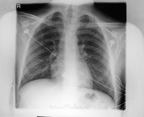
Subpulmonic effusion Normal Chest
Note the flat looking "hemidiaphragm", particularly medially. When you compare this appearance with the normal chest, the differences may appear subtle. I would argue that the fact that the patient has a raised right hemidiaphragm, the flattened look of the diaphragm and the concave looking horizontal fissure would be enough to suggest a subpulmonic effusion
<a class="external" href="http://www.med.yale.edu/intmed/cardio/imaging/cases/pleural_effusion_subpulmonic/index.html" rel="nofollow" target="_blank">http://www.med.yale.edu/intmed/cardio/imaging/cases/pleural_effusion_subpulmonic/index.html</a>
Compare this normal right hemidiaphragm shape to that of the patient with the right sided subpulmonic effusion.
A subpulmonic effusion can be mimicked by "subdiaphragmatic abscess, enlarged liver, paralysis and eventration of the diaphragm, and ascites" (2)
Case 1
| | |
|
| | |
|
Case 2
| | |
|
| | |
|
Discussion
ReferencesNot every patient who has a subpulmonic effusion warrants a lateral decubitus chest view. It would not be unusual, for example, for a patient to have a subpulmonic effusion post thoracic surgery. Subjecting a patient to the rigors of adopting the lateral decubitus position unnecessarily is undesirable for a variety of reasons.
Where a subpulmonic effusion is suspected on the PA chest image, and is likely to be a significant finding, a lateral decubitus chest view may assist the radiologist, referring doctor and ultimately the patient.
1. MI Schwarz and BL Marmorstein A new radiologic sign of subpulmonic effusion. <a class="external" href="http://www.chestjournal.org/cgi/reprint/67/2/176.pdf" rel="nofollow" target="_blank">http://www.chestjournal.org/cgi/reprint/67/2/176.pdf</a>
http://chestjournal.org/cgi/content/abstract/67/2/176
2. Rothstein E, Landis FB: Infrapulmonary pleural effusion simulating elevation of the hemidiaphragm. Am J Med 8:4852, 1950
...back to the Applied Radiography home page
