Gallstones
TerminologyThe imaging of gallstones has undergone considerable change over the last 30 years. The procedure of oral cholecystography has been superseded by ultrasound imaging. The plain film appearance of gallstones is worthy of consideration given its relatively common occurrence and its potential to account for patient symptoms. Furthermore, demonstration of gallstones on plain film can be a noteworthy incidental finding.
Presence of stones in the gallbladder is referred to as cholelithiasis
Choledocholithiasis is the presence of gallstones in the common bile duct.
Acute cholangitis is a bacterial infection superimposed on an obstruction of the biliary tree most commonly from a gallstone
Cholecystitis is inflammation of the gallbladder, usually resulting from a gallstone blocking the cystic duct
Pathophysiology and Gallstone Types
Gallstone formation occurs because certain substances in bile are present in concentrations that approach the limits of their solubility. When bile is concentrated in the gallbladder, it can become supersaturated with these substances, which then precipitate from solution as microscopic crystals. The crystals are trapped in gallbladder mucus, producing gallbladder sludge. Over time, the crystals grow, aggregate, and fuse to form macroscopic stones. Occlusion of the ducts by sludge and stones produces the complications of gallstone disease.The 2 main substances involved in gallstone formation are cholesterol and calcium bilirubinate.
•Stone formation is based on the relative concentration of cholesterol, bile salts, and phospholipid
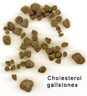
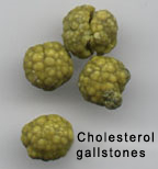
Gallstone Type Notes Appearance cholesterol More than 80% of gallstones in the United States contain cholesterol as their major component yellow
<a class="external" href="http://www.gihealth.com/html/education/photo/gallstones.html" rel="nofollow" target="_blank">http://www.gihealth.com/html/education/photo/gallstones.html</a>calcium bilirubinate Bilirubin, a yellow pigment derived from the breakdown of heme, is actively secreted into bile by liver cells
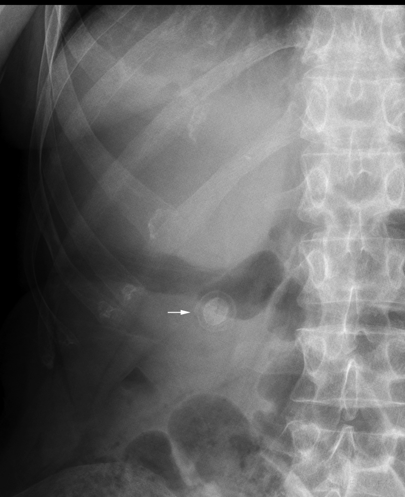
10-20% of gallstones in the United Statesgreen- brown Mixed Cholesterol gallstones may become colonized with bacteria and can elicit gallbladder mucosal inflammation. Lytic enzymes from bacteria and leukocytes hydrolyze bilirubin conjugates and fatty acids. As a result, over time, cholesterol stones may accumulate a substantial proportion of calcium bilirubinate and other calcium salts, producing mixed gallstones. Large stones may develop a surface rim of calcium resembling an eggshell that may be visible on plain x-ray films.
Treatment<a class="external" href="http://emedicine.medscape.com/article/175667-overview" rel="nofollow" target="_blank">Douglas M Heuman, Cholelithiasis</a>
Risk Factors
- Laparoscopic cholecystectomy
- lithotripsy
Whilst there is a long list of risk factors for gallstone formation, the commonly remembered and frequently cited mnemonic is the 4 "F"s
- female
- fat
- fertile and
- forty
Radiological Appearance
only 15% of gallstones are radiopaque
Solitary layered gallstone arrowed
Case 1
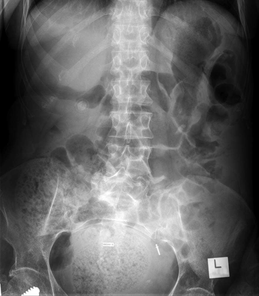
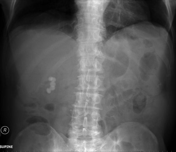
This 89 year old male presented to the Emergency Department with symptoms consistent with cholecystitis. He was referred for an abdominal plain film.
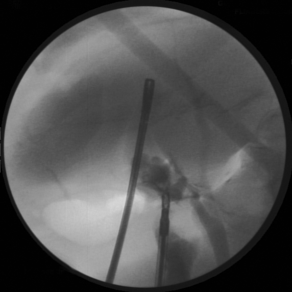
There appeared to be 5 well defined radiopaque gallstones in the RUQ.The patient underwent laparoscopic cholecystectomy with laparascopic cholangiography. The patient developed some post op abdominal discomfort and was referred for an abdominal plain film.
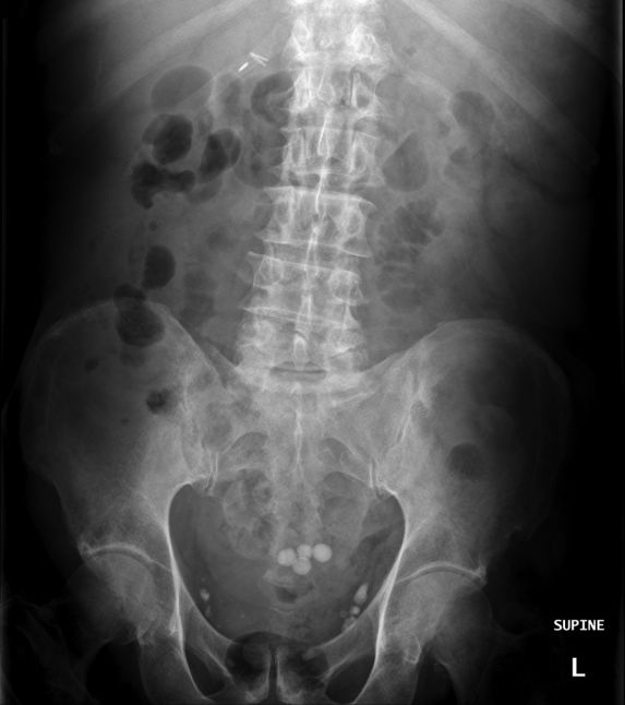
4 gallstones were demonstrated overlying the sacrum
Metallic sutures noted in RUQ associated with recent laparscopic cholecystectomy
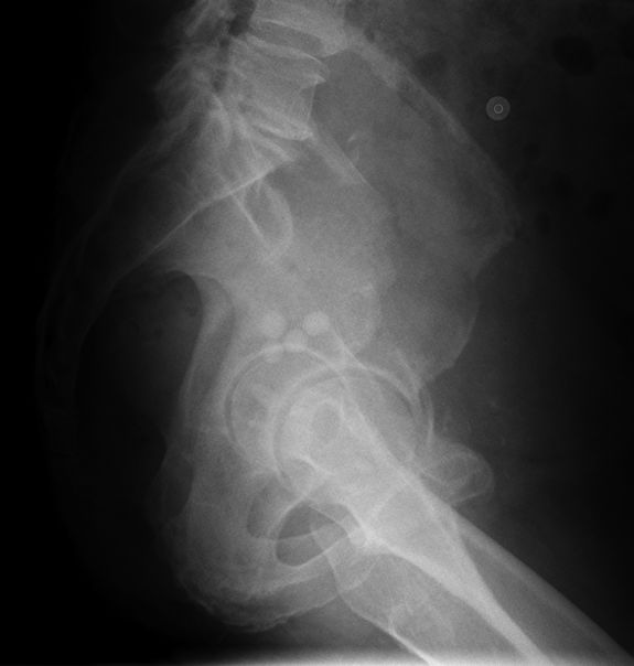
Phlebelihs also notedFurther abdominal plain film imaging was requested to establish whether the stones were likely to be sited within the pouch of Douglas.
It appeared likely that the the gallstones had descended to the most dependent portion of the pelvic peritoneal cavity or pouch of Douglas
The pouch of Douglas (cul-de-sac) represents the caudal extension of the peritoneal cavity. It is the rectovaginal pouch in the female and the rectovesical pouch in the male. The cul-de-sac is in a dependent position when either upright or supine; it is, therefore, a frequent location for seeded lesions.
<a class="external" href="http://www.ncbi.nlm.nih.gov/pubmed/3053046" rel="nofollow" target="_blank">http://www.ncbi.nlm.nih.gov/pubmed/3053046</a>
... back to the Wikiradiography home page
... back to the Applied Radiography home page
