DISI and VISI Deformities
The recognition of DISI (dorsal intercalated segmental instability) and VISI (Volar intercalated segmental instability) on plain film images of the wrist is within the pattern recognition skills of trauma radiographers who are operating at an advanced level. It is important that standard views of the wrist (particularly the lateral view) are performed as supplementary views when a DISI or VISI is suspected..
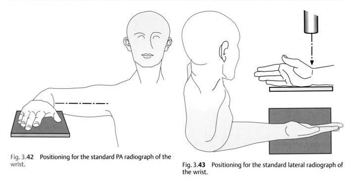
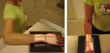
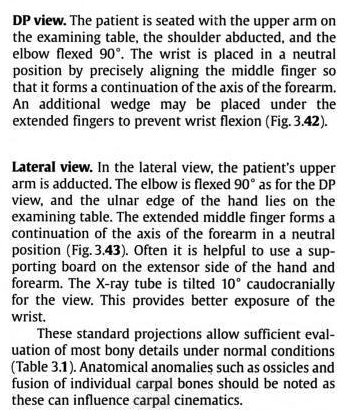
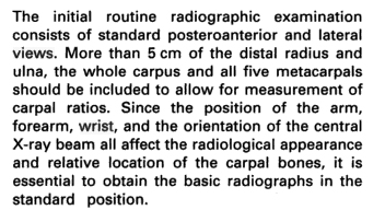
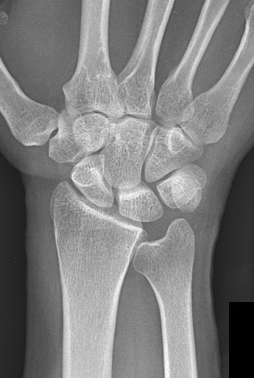
Standard Views of the Wrist
Comment
These descriptions are from radiology-based texts rather than radiography texts. The radiography technique textbooks do not consider the position of the forearm/elbow when describing wrist positioning. The positions as described above are akin to 'mixed forearm' views which are performed when patients have limited movement in their arm (usually from pain) or when the patient has an above elbow cast. The wrist positioning approach described above will tend to produce two views of the anatomy at 90 degrees because the relationship between the radius and ulna does not change- the 90 degree movement occurs largely at the shoulder.
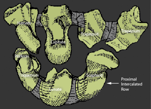
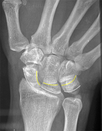
The Intercalated Row Concept
adapted from <a class="external" href="http://www.orthospot.com.au/papers.orthospot.com.au/fracupl_files/frame.htm" rel="nofollow" target="_blank">http://www.orthospot.com.au/papers.orthospot.com.au/fracupl_files/frame.htm</a>"The row concept best explains the behaviour of the carpal bones. The proximal row acts as an intercalated segment- there is no direct control of this row.
The lunate is like the hooker of a rugby scrum. It is controlled by the bones surrounding it via the short interosseous ligaments. It has a natural tendency if isolated to 'pop out' and tilt dorsally."
quoted from <a class="external" href="http://www.orthospot.com.au/papers.orthospot.com.au/fracupl_files/frame.htm" rel="nofollow" target="_blank">http://www.orthospot.com.au/papers.orthospot.com.au/fracupl_files/frame.htm</a>
DISI and VISI Plain Film Signs
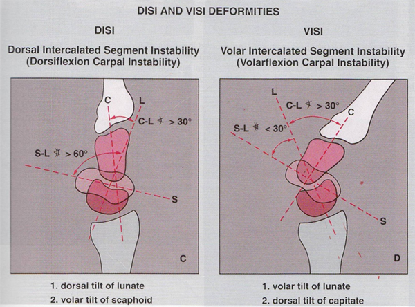
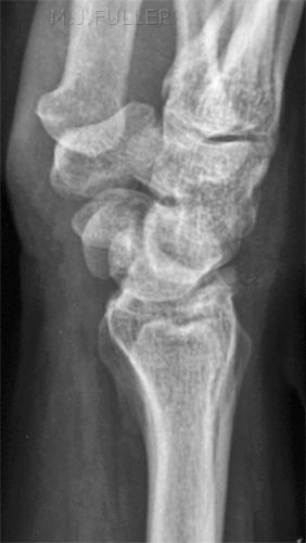
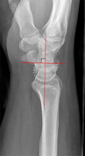
<a class="external" href="http://books.google.com.au/books?id=frtqnmt0lDoC&pg=PA206&dq=disi+visi" rel="nofollow" target="_blank">Adam Greenspan. Orthopedic Imaging: A Practical. Approach, 4th edition. Philadelphia, Pa: Lippincott Williams. & Wilkins, 2004. p206</a>Don't be put-off by the apparent complexity of the deformities. A simple approach is to consider the lunate which is usually the easiest carpal bone to visualise on a lateral wrist image. If the lunate is abnormally tilted in a dorsal direction on a standard lateral wrist image, a DISI should be considered. If the lunate is abnormally tilted in a volar direction a VISI should be considered.
DISI (dorsal intercalated segmental instability)
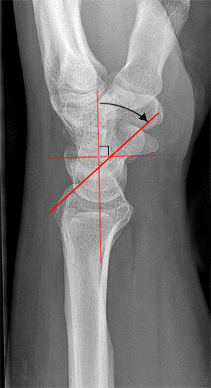
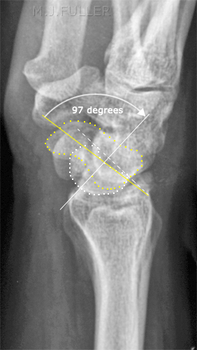
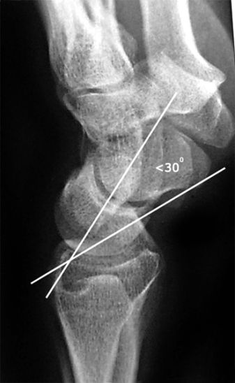
note that this angle has a larger than normal error because the wrist is not in a true lateral positionDISI, or dorsiflexion instability, is short for
dorsal intercalated segmental instability.
The intercalated segment is the proximal carpal row identified by the lunate. The term 'intercalated segment' refers to it being the part in between the proximal segment of the wrist consisting of the radius and the ulna and the distal segment, represented by the distal carpal row and the metacarpals.
So all this means is that in DISI, or dorsiflexion instability, the lunate is angulated dorsally.
If you think the lunate is tilted, measure the scapholunate angle ( 30-60°is normal, 60-80°is questionably abnormal, >80° is abnormal) and the capitolunate angle (<30° is normal).
quoted from<a class="external" href="http://www.radiologyassistant.nl/en/42a29ec06b9e8" rel="nofollow" target="_blank">Louis A. Gilula and Ileana Chesaru</a>
<a class="external" href="http://www.radiologyassistant.nl/en/42a29ec06b9e8" rel="nofollow" target="_blank">Wrist - Carpal instability
The Radiology Assistant</a>
DISI is due to disruption of the scapho-lunate articulation
A DISI will typically demonstrate a scapho-lunate angle greater than 60 degrees
quoted from
<a class="external" href="http://books.google.com.au/books?id=qTCbB2N2UFcC&pg=PA54&lpg=PA54&dq=disi+visi&source=bl&ots=jrX-iBQZ9o&sig=ueTw6hUN5wk__z_SNTKm_3X0V6A&hl=en&ei=ikgTSvaNCJS-tAOy68XdDQ&sa=X&oi=book_result&ct=result&resnum=4#PPA55,M1" rel="nofollow" target="_blank">Giuseppe Guglielmi, Cornelis Van Kuijk, Harry K. Genant, Fundamentals of hand and wrist imaging, 2001</a>
DISI pattern:adapted from <a class="external" href="http://som.flinders.edu.au/FUSA/ORTHOWEB/notebook/regional/wrist.html" rel="nofollow" target="_blank">http://som.flinders.edu.au/FUSA/ORTHOWEB/notebook/regional/wrist.html</a>
- when the scapholunate joint is dissociated, the scaphoid is palmar flexed and the lunate is dorsiflexed
- Scapho-lunate angle usually 30- 60degrees (average 46 degrees) and with DISI it is greater than 70degrees
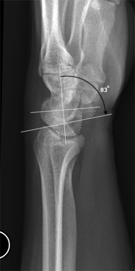
Case Study
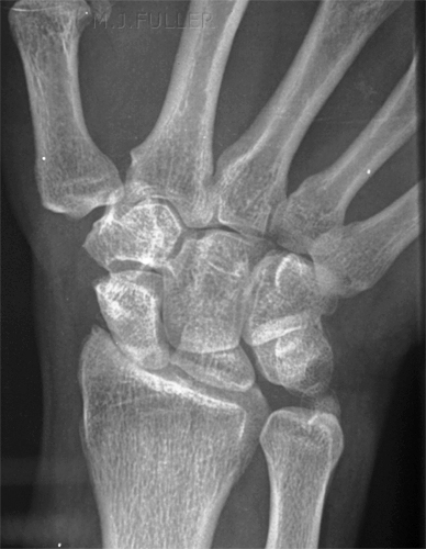
This patient presented to the Emergency Department following a fall from 3 metres. He had severe pain in his right wrist.
VISI (Volar intercalated segmental instability)
VISIadapted from
<a class="external" href="http://imaging.birjournals.org/cgi/content/full/15/4/180" rel="nofollow" target="_blank">P S McAlinden and J Teh,
</a><a class="external" href="http://imaging.birjournals.org/cgi/content/full/15/4/180" rel="nofollow" target="_blank">Imaging of the wrist, Imaging 15:180-192 (2003)</a>
While most DISI is abnormal, in many cases VISI is a normal variant, especially if the wrist is very lax.
quoted from<a class="external" href="http://www.radiologyassistant.nl/en/42a29ec06b9e8" rel="nofollow" target="_blank">Louis A. Gilula and Ileana Chesaru</a>
<a class="external" href="http://www.radiologyassistant.nl/en/42a29ec06b9e8" rel="nofollow" target="_blank">Wrist - Carpal instability
The Radiology Assistant</a>
VISI is secondary to disruption of the lunate and triquetral
quoted from
<a class="external" href="http://books.google.com.au/books?id=qTCbB2N2UFcC&pg=PA54&lpg=PA54&dq=disi+visi&source=bl&ots=jrX-iBQZ9o&sig=ueTw6hUN5wk__z_SNTKm_3X0V6A&hl=en&ei=ikgTSvaNCJS-tAOy68XdDQ&sa=X&oi=book_result&ct=result&resnum=4#PPA55,M1" rel="nofollow" target="_blank">Giuseppe Guglielmi, Cornelis Van Kuijk, Harry K. Genant, Fundamentals of hand and wrist imaging, 2001</a>
VISI pattern:adapted from <a class="external" href="http://som.flinders.edu.au/FUSA/ORTHOWEB/notebook/regional/wrist.html" rel="nofollow" target="_blank">http://som.flinders.edu.au/FUSA/ORTHOWEB/notebook/regional/wrist.html</a>
- lunate palmar flexed
- if the lunate and triquetrum can be seen, the normal lunotriquetral angle of approximately -16 degrees becomes neutral or positive
Comment
Carpal ligament injuries are easily missed on plain film images. Patients with these injuries are at risk of misdiagnosis and inappropriate treatment. Radiographers who are able to recognise carpal bone deformities associated with ligament rupture are in a position to perform appropriate supplementary views. If you are able to recognise these types of carpal bone deformities, you can consider yourself to be operating at a very advanced level as a trauma radiographer.
...back to the Wikiradiography home page
...back to the Applied Radiography home page
