Clavicle Radiography
For those radiographers who are employed in facilities that provide trauma and acute care services, clavicle fractures are commonplace. This page considers clavicle radiography techniques and clavicle trauma image interpretation.
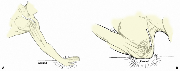
Mechanism of Injury
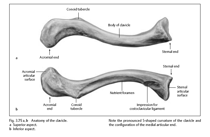
Anatomy
<a class="external" href="http://www.buchhandel.de/WebApi1/GetMmo.asp?MmoId=2998578&mmoType=PDF" rel="nofollow" target="_blank">Source: Koehler/Zimmer's Borderlands of Normal and Early Pathological Findings in Skeletal Radiography, Thieme, 2003, 5th Edition, p306</a>This image is taken from a book titled
<a class="external" href="http://www.buchhandel.de/WebApi1/GetMmo.asp?MmoId=2998578&mmoType=PDF" rel="nofollow" target="_blank">Borderlands of Normal and Early Pathological Findings in Skeletal Radiography</a>. This is a textbook with some unique information that is well worth investing in.
<embed allowfullscreen="true" height="350" src="http://widget.wetpaintserv.us/wiki/wikiradiography/widget/youtubevideo/c9aee36f6f06af2b80e71f9059a2e1b2949b9a74" type="application/x-shockwave-flash" width="425" wmode="transparent"/> <embed allowfullscreen="true" height="350" src="http://widget.wetpaintserv.us/wiki/wikiradiography/widget/youtubevideo/68ab6a6b0a980ba1eb39ea31e9defcb2886d3288" type="application/x-shockwave-flash" width="425" wmode="transparent"/> <embed allowfullscreen="true" height="350" src="http://widget.wetpaintserv.us/wiki/wikiradiography/widget/youtubevideo/e57877d96651b712e1ba027a6f0ba52fb78244e7" type="application/x-shockwave-flash" width="425" wmode="transparent"/>
<a class="external" href="http://www.buchhandel.de/WebApi1/GetMmo.asp?MmoId=2998578&mmoType=PDF" rel="nofollow" target="_blank">Source: Koehler/Zimmer's Borderlands of Normal and Early Pathological Findings in Skeletal Radiography, Thieme, 2003, 5th Edition, p307</a>
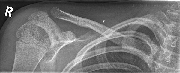
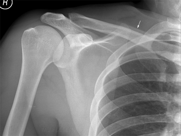
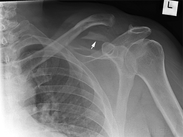
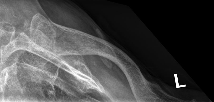
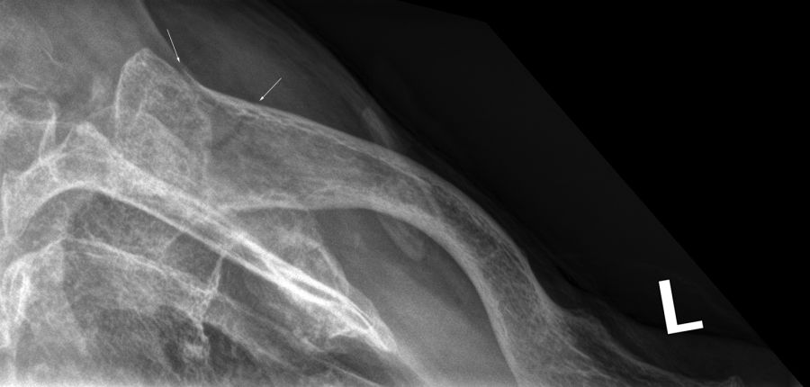
The arrowed sructure is commonly referred to as a clavicle companion shadow. This line is caused by the skin and subcutaneous tissues curving around the clavicle.
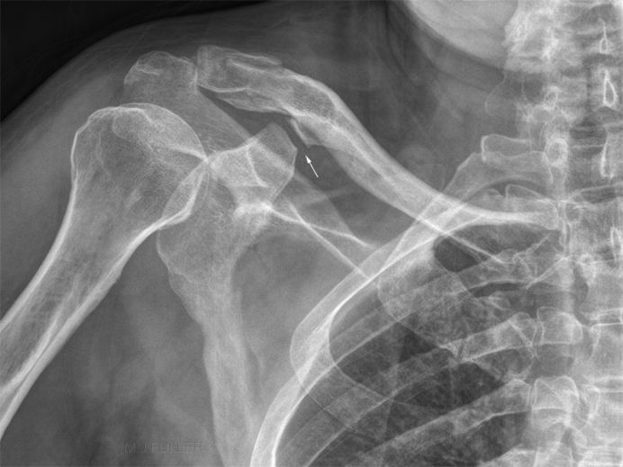
The arrowed structure is referred to as a coracoclavicular joint. This is an anomalous articulation between the coracoid and the clavicle.
Clinical Presentation
Patients with clavicle fracture will almost invariably be experiencing pain associated with the fracture. Given that the clavicle is a superficial bony structure, a clinical diagnosis is often made with confidence. Consideration of acromioclavicular joint injury and sternoclavicular joint injury should be made, particularly in patients who have significant symptoms (pain) and do not demonstrate a clavicle fracture radiographically.
The following images follow a clavicle fracture from initial presentaion to post-op imaging.
Andrew H. Schmidt, M.D., T.J. McElroy, January 2007
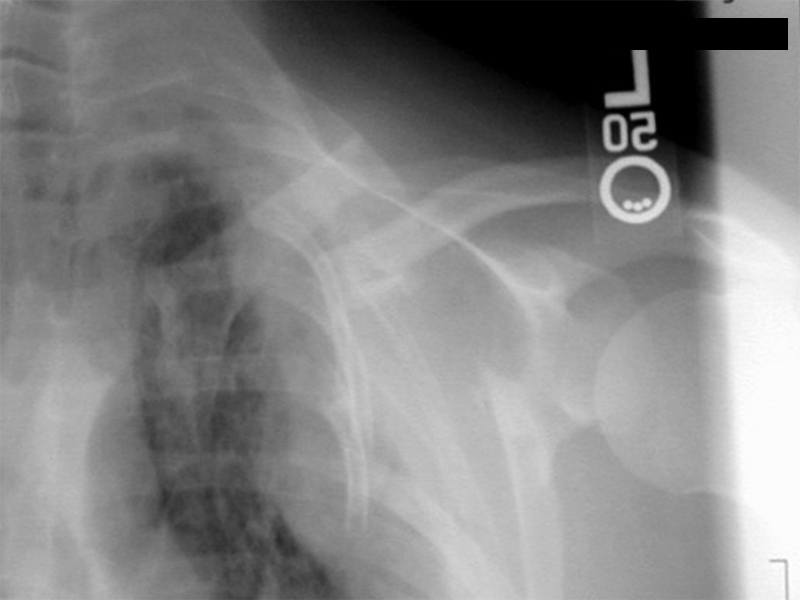
U01 clavicle AC SC Joints 1.pptThis patient has suffered considerable trauma resulting in left sided scapula fracture, multiple rib fractures and clavicle fracture.
The arrows indicate the overlapping segments of the fractured clavicle
Andrew H. Schmidt, M.D., T.J. McElroy, January 2007
U01 clavicle AC SC Joints 1.pptThe AP shoulder image demonstrates a mid-clavicle fracture, fractured scapula, haemothorax and multiple rib fractures (? flail segment)
Andrew H. Schmidt, M.D., T.J. McElroy, January 2007
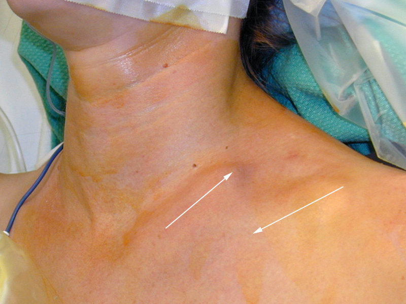
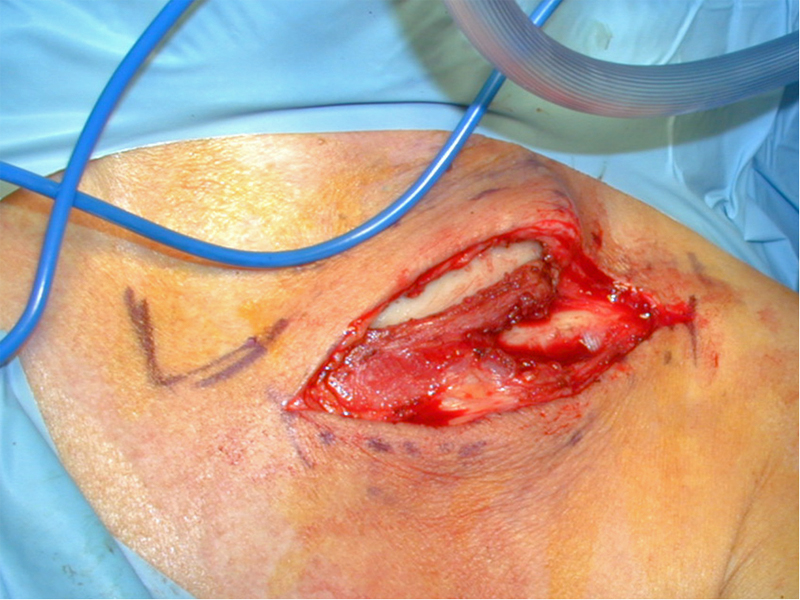
U01 clavicle AC SC Joints 1.pptThe clavicle fracture exposed at surgery
Andrew H. Schmidt, M.D., T.J. McElroy, January 2007
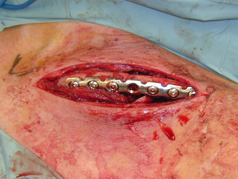
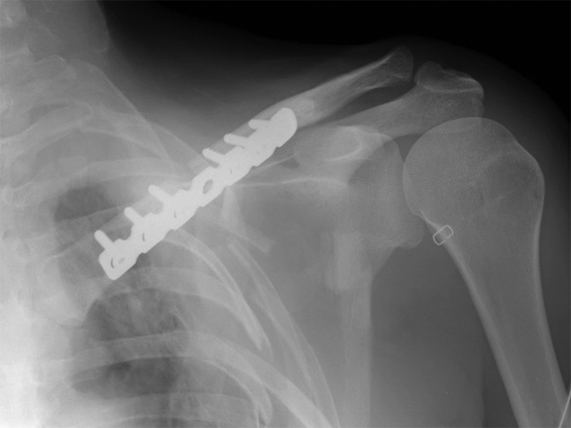
U01 clavicle AC SC Joints 1.pptThe ORIF of the left clavicle fracture with screws and plate.
Radiography
Andrew H. Schmidt, M.D., T.J. McElroy, January 2007
<a class="external" href="http://www.scribd.com/doc/21709781/U01-Clavicle-AC-SC-Joints" rel="nofollow" target="_blank">U01 clavicle AC SC Joints 1.ppt</a>Post operative radiography
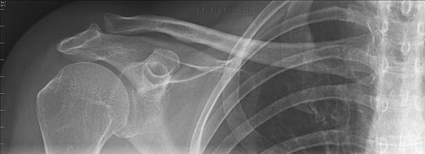
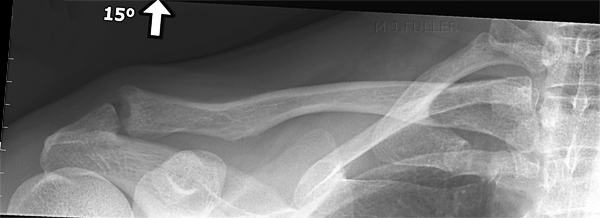
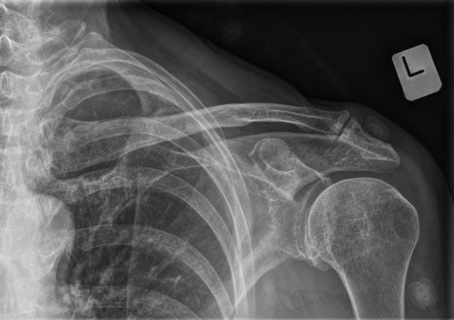
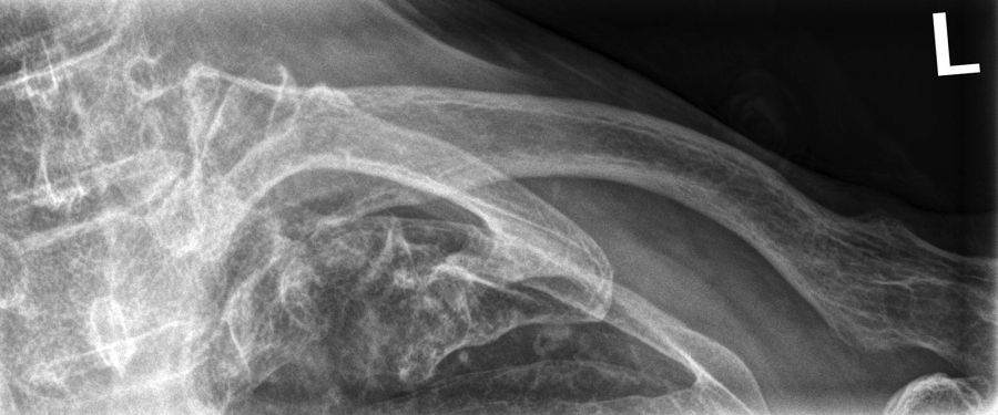
A typical trauma radiography series of the clavicle will include an AP shoulder projection and an AP clavicle projection with cephalic angulation. This series is most suitable for patients who present with convincing clinical signs of an isolated clavicle fracture. The AP shoulder projection is useful in diagnosing other (unsuspected) shoulder girdle bony injury. The cephalic angle clavicle projection should be included to avoid missing subtle clavicle fractures. This view also has the potential to provide an improved appreciation of the nature of the fracture and its degree of displacement. See cases below which illustrate these points.
<a class="external" href="http://books.google.com.au/books?id=7ca8iqAPo2UC&pg=PA597&lpg=PA597&dq=clavicle+radiography&source=bl&ots=Wk8S-xPBA9&sig=CjJ3MqglNxpBKE26nynuOgva-8c&hl=en&ei=IRMjStShIpOVkAXMidSVBQ&sa=X&oi=book_result&ct=result&resnum=17#PPA597,M1" rel="nofollow" target="_blank">Charles A. Rockwood, Frederick A. Matsen, III, Michael A. Wirth, Steven B. Lippitt.
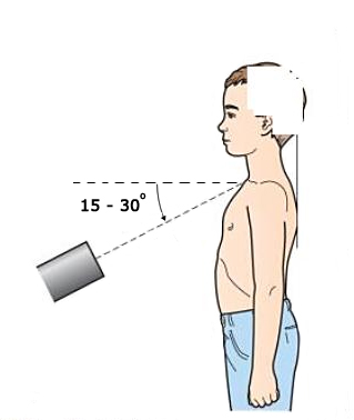
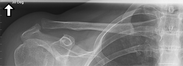
The Shoulder, 2009</a>Clavicle radiography is frequently performed with the patient in the AP erect position as shown in this illustration. There are multiple variations on this technique including PA imaging and patient angulation rather than tube angulation. This is an AP projection clavicle image with no tube or patient angulation. This is an AP projection of the clavicle with 15 degrees of cephalic tube angulation. The tube angulation can typically range from 15 to 30 degrees. The greater the tube angulation applied, the greater the superior projection of the clavicle.
Inclusion of the SC joint is reasonable in trauma situations.Note that the AC joint is more clearly demonstrated using this projection compared to the straight tube (non-angled) AP projection of the clavicle.
Pathology
<a class="external" href="http://www.boddunan.com/education/20-medicine-a-surgery/3528-sternocleidomastoid.html" rel="nofollow" target="_blank">
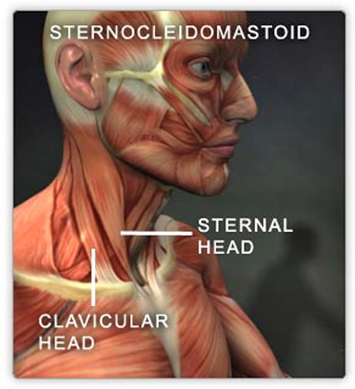
http://www.boddunan.com/education/20-medicine-a-surgery/3528-sternocleidomastoid.html</a>Displacement of the clavicular fracture is always as shown- the Sternocleidomastoid muscle tends to pull the proximal fragment in a cranial direction and gravity largely takes care of the distal fragment. Sometimes there is a comminuted fragment almost turned at right angles between the ends.
Case Studies
Case 1
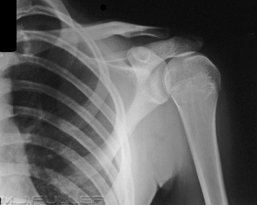
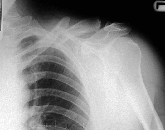
This patient presented to the Emergency Department following a fall. The patient underwent a clinical assessment and was subsequently referred for shoulder radiography with a clinical diagnosis of clavicle fracture.
It is apparent that there is no clavicle fracture. The clavicle appears to have smooth bony contours and there is no obvious soft tissue swelling- the soft tissue line visualized running parallel to the upper border of the clavicle is known as a companion shadow and appears unremarkable. At this point it would be reasonable to question the value of any further imaging of the patient’s clavicle. Further imaging of the clavicle would appear to be contributing to the patient’s radiation dose without adding any diagnostic value.
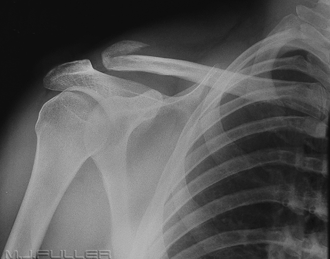
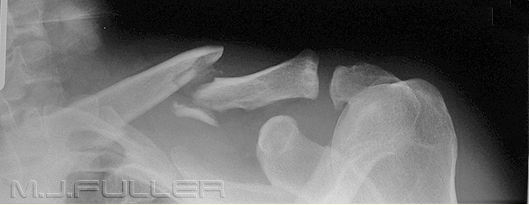
The radiographer continued the examination by performing a dedicated collimated AP cephalic angle clavicle projection as shown below-left.There is clearly a midshaft clavicle fracture that was not demonstrated on the first image This fracture was superimposed over the patient’s second rib causing it to be completely obscured. In retrospect, there may have been minimal distortion of the clavicle companion shadow on the initial image, but this could have easily have been overlooked.
Case 2
Case 3
Case 4
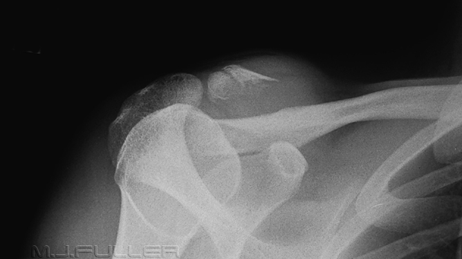
CommentThis case presents a good example of a clinical approach to radiography. The radiographic series was supplemented with additional projections to demonstrate the fracture that the radiographer considered to be clinically evident.
Summary
Relevant wikiRadiography LinksThe temptation not to perform a dedicated cephalic angle clavicle projection can be overwhelming in cases where you think that you have clearly demonstrated, or not demonstrated, a clavicle fracture on the AP shoulder image. However, it comes with a diagnostic risk. Single view radiography of any bony structure is a hazardous practice and should be avoided whenever possible. In the words of John Harris, "one view is no view".
....back to the applied radiography home page here
