Calcaneal Fractures
Calcaneal injuries are one of the less common injuries seen in acute care settings (compare the number of calcaneal fractures you see with the number of ankle fractures) . An awareness of normal anatomical appearances, normal anatomical variants, common pathologies and common pitfalls will assist the radiographer to avoid misdiagnosis.
Mechanisms of Injury
Fall from a height is the most common cause of calcaneum fracture. This type of a calcaneal fracture is known as a Lover's fractures, Don Juan fracture or Casanova Fracture (based on sequelae of jumping from a bedroom window onto a hard surface).
Associated Fractures
Anatomy and Normal Anatomical VariantsConsideration should be given to the possibility that a fractured calcaneum might be bilateral. A fracture of the calcaneum is also associated with thoraco-lumbar fractures.
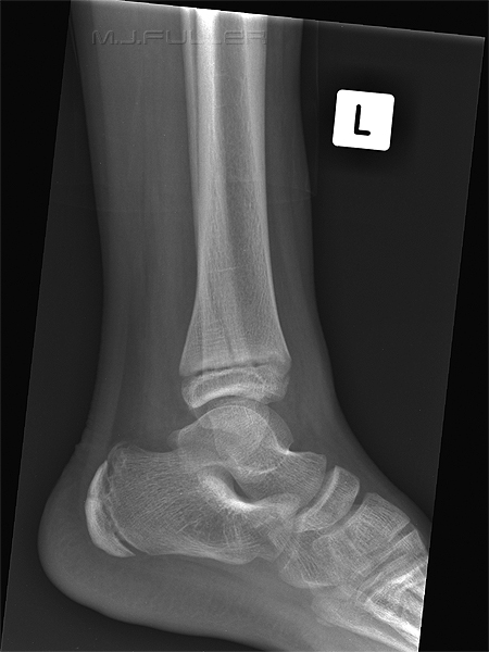
This is the calcaneum of a child demonstrating the normal calcaneal apophysis. This would not normally cause diagnostic confusion.
Radiographic Technique
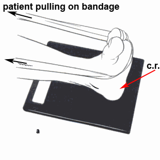
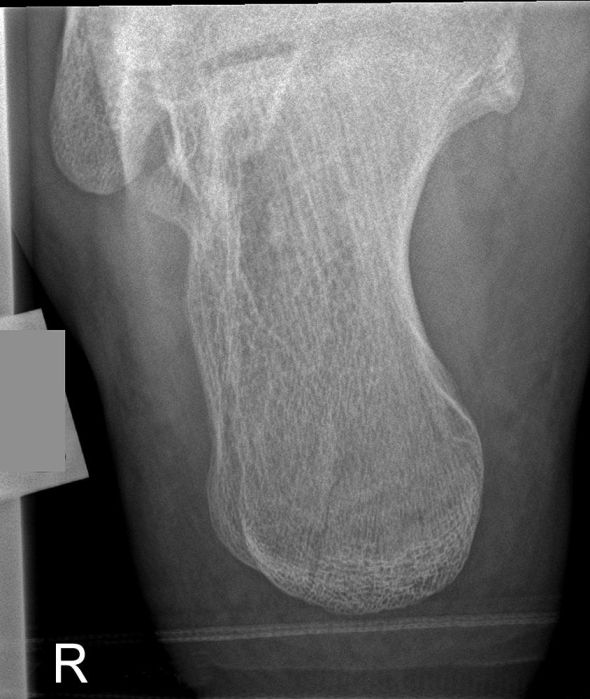
adapted from <a class="external" href="http://books.google.com.au/books?id=NePk5A1Y1NAC&printsec=frontcover" rel="nofollow" target="_blank">Skeletal Radiography: A Concise Introduction to Projection Radiography</a><a class="external" href="http://books.google.com.au/books?id=NePk5A1Y1NAC&printsec=frontcover" rel="nofollow" target="_blank">By Sheila Bull</a>The axial view of the calcaneum is the one that causes problems. Ideally, the patient should dorsiflex their foot -you would ask the patient to "bring your toes toward you". Of course, this is not likely to happen if the patient has a significant calcaneum injury- it's simply too painful. The tricky part is knowing how much cephalic tube angle to use. The textbooks say 40 degrees, but most experienced radiographers make a judgement based on how much the patient can dorsiflex their foot whilst taking into consideration that you do not want to end up with an extremely elongated image of the calcaneum.
If the patient is not in too much pain, a bandage can be wrapped around the foot with the patient pulling on the bandage to achieve maximum dorsiflexion of the ankle. This would not typically be a trauma technique.
adapted from
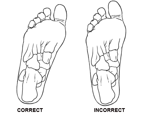
<a class="external" href="http://books.google.com.au/books?id=NePk5A1Y1NAC&printsec=frontcover" rel="nofollow" target="_blank">Skeletal Radiography: A Concise Introduction to Projection Radiography</a><a class="external" href="http://books.google.com.au/books?id=NePk5A1Y1NAC&printsec=frontcover" rel="nofollow" target="_blank">By Sheila Bull</a>Internal rotation of the foot will improve the appearance of the calcaneum in the axial projection by orientating the calcaneum in a vertical position. (I could not find this mentioned in any of the textbooks, perhaps it is just one of my quirks)
Kager's Fat Pad
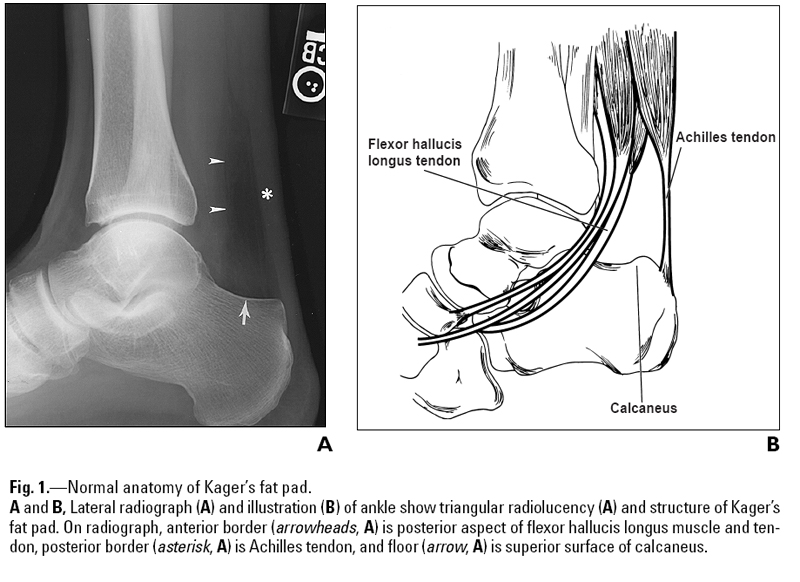
<a class="external" href="http://www.ajronline.org/cgi/content/full/182/1/147" rel="nofollow" target="_blank">Justin Q. Ly and Liem T. Bui-Mansfield Anatomy of and Abnormalities Associated with
Kager’s Fat Pad AJR:182, January 2004</a>It is not uncommon for ankle and calcaneal injuries to involve Kager's fat pad. A careful examination of the density, shape and borders of Kager's fat pad can provide indicators of bony injury to the ankle and calcaneum. An abnormal Kager's fat pad does not indicate definite bony injury to the ankle, rather, it indicates that a close examination of the anatomy is warranted and may lower your threshold for undertaking supplementary views.
Boehler’s Angle (aka Bohler's Angle)
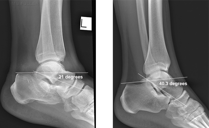
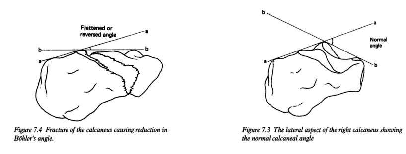
"Subtle fractures may only be identified by assessing Boehler’s angle. This angle is measured by drawing a line from the highest point of the posterior tuberosity to the highest midpoint, and a 2nd line from the highest midpoint to the highest point of the anterior process. The angle, posteriorly, should be >30 degrees. If there is flattening of the bone due to a fracture, this angle will be decreased, to <30 degrees." <a class="external" href="http://imageinterpretation.co.uk/ankle.html" rel="nofollow" target="_blank">http://imageinterpretation.co.uk/ankle.html</a>
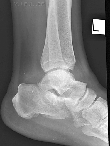
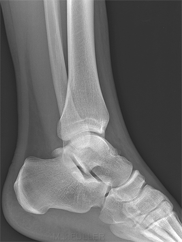
adapted from<a class="external" href="http://books.google.com.au/books?id=NePk5A1Y1NAC&printsec=frontcover" rel="nofollow" target="_blank">Skeletal Radiography: A Concise Introduction to Projection Radiography</a><a class="external" href="http://books.google.com.au/books?id=NePk5A1Y1NAC&printsec=frontcover" rel="nofollow" target="_blank">By Sheila Bull</a>The first image demonstrates a Boehler's Angle of 21 degrees suggesting that there is a fracture of the calcaneum. Compare this with the normal anatomy on the right
The Value of the Axial View of the Calcaneum
Case 1
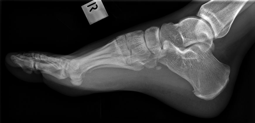
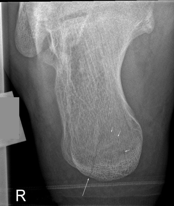
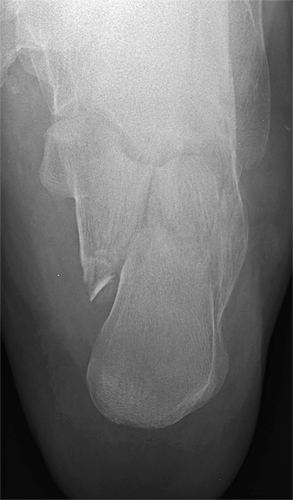
The lateral view of the calcaneum is easier to achieve than the axial view. It can be tempting to not perform the axial view when the lateral view reveals no bony abnormality. This practice courts misdiagnosis- I have seen several cases where the axial view demonstrated a calcaneal fracture that was not apparent on the lateral view.
CommentIf the mechanism of injury and/or the clinical signs suggest the possibility of calcaneal fracture, an axial calcaneal projection must be included in the series. Missed calcaneal fractures could result in patient's suffering considerable pain and discomfort as they attempt to mobilise on their fractured calcaneum.
Case 2
back to the Applied Radiography home page
