Bone Tumours
Introduction
Bone tumours and tumour-like lesions can present a challenge for both radiographer and radiologist. This page is an alphabetical listing of bone tumours and tumour-like lesions.
Enchondroma
Fibrous Cortical Defect
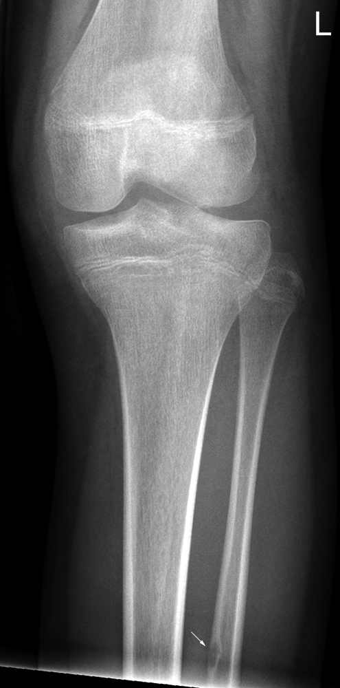
This 12 year old girl was referred for knee radiography post arthroscopy. A fibrous cortical defect of the fibula (arrowed) was noted as an incidental finding.
Fibrous cortical defects are benign lesions commonly found as an incidental finding in the metaphysis of long bones in children.A fibrous cortical defects typically arises in the metaphyses of long bones particularly the distal femur and tibia. It is common, possibly affecting more than 40% of boys and 30% of girls with an age range 2 - 15 years. The lesion is thought to originate at the insertion site of a ligament or tendon and it has been suggested that it may reflect a previous traction injury. Although the lesion arises in the metaphyseal region it may migrate towards the diaphysis with growth. The lesions are usually asymptomatic, although they are occasionally associated with pathological fractures.
Radiographs show an eccentric lucent lesion with thinned cortex, which may have a multilocular appearance and often a sclerotic margin. The lesions spontaneously regress with time; radiographs will show increasing marginal sclerosis followed by progressive ossification of the lesion extending from its diaphyseal aspect. The appearance on conventional radiographs should be characteristic and no further imaging is indicated. However, the lesions are commonly seen as incidental findings on other imaging. Scintigraphy may show increased activity depending on the stage of healing. Likewise MR imaging may show variable signal intensity depending the lesion's stage of healing. There is often central decreased T2-weighted signal due to collagen and haeomosiderin deposition.
Lesions persisting in older children which are more than 2 cm in size have been termed nonossifying fibromas but appear to represent the same histological entity. They often extend into the medullary cavity and are associated with an increased risk of pathological fracture due to their size.
<a class="external" href="http://www.medcyclopaedia.com/library/topics/volume_vii/f/fibrous_cortical_defect/gfibrous_cortical_defect_fig1.aspx" rel="nofollow" target="_blank">medcyclopaedia</a>
Non-Ossifying Fibroma (Fibroxanthoma)
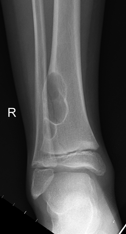
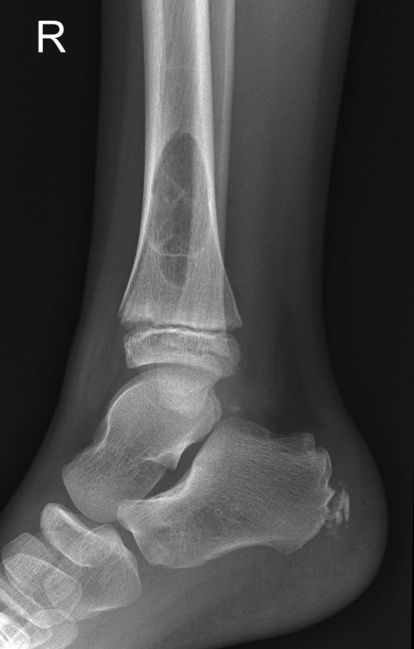
AP ankle image of a 10year old boy who presented to the Emergency Department with a painful ankle following minor trauma. The distal tibia shows a lobulated circumscribed non-ossifying fibroma that is eccentrically located within the distal tibial metadiaphysis. Peripheral sclerotic border with a central lucency is typical of this lesion. There is a Salter Harris II pathological fracture associated with this lesion.
(CR screen cleaning artifact noted)
<a class="external" href="http://emedicine.medscape.com/article/389590-overview" rel="nofollow" target="_blank">
</a>
Discussion
Fibroxanthoma is the preferred term for the non-ossifying fibroma (NOF) lesion because it more accurately reflects the underlying pathologic findings. <a class="external" href="http://emedicine.medscape.com/article/389590-overview" rel="nofollow" target="_blank">(Stacy E Smith, Fibrous Cortical Defect and Nonossifying Fibroma, e-medicine)</a>. NOF/fibroxanthoma is one of the more commonly seen bone lesions. There is some debate regarding the distinction between NOF/fibroxanthoma and fibrous cortical defect. Some authors have argued that NOF/fibroxanthoma and fibrous cortical defects can be distinguished by their "...size and natural history" <a class="external" href="http://emedicine.medscape.com/article/389590-overview" rel="nofollow" target="_blank">(Stacy E Smith, Fibrous Cortical Defect and Nonossifying Fibroma, e-medicine)</a>. Clyde Helms argues that the two lesions are histologically identical and "... it is illogical to subdivide the lesion by its size" (Clyde Helms, Fundamentals of Skeletal Radiology, 3rd ed, Elsevier, Philadelphia 2005, p16).
NOF/fibroxanthoma are relatively commonly seen in children and tend to regress before aldulthood. Clyde Helm's describes non-ossifying fibromas as follows
"NOF's are benign, asymptomatic lesions that typically occur in the metaphysis of a longbone emanating from the cortex...They classically have a thin, sclerotic border that is scalloped and slightly expansile; however, this is a general description that applies to only 75% of lesions. They do not have to have expansion or a scalloped or sclerotic border, and they are not limited to the metaphysis.
... These lesions are so characteristic that no lgical differential diagnosis should be entertained..If the patient is older than 30 years I will not include NOF in the differential diagnosis."
Clyde Helms, Fundamentals of Skeletal Radiology, 3rd ed, Elsevier, Philadelphia 2005, p16
At [[1]] the following features of NOF are noted
- Most often occur in lower extremities around knee
- Fewer than 10% occur in upper extremities
- Characteristics
- Geographic
- Lytic
- Multilobulated
- Metaphyseal
- Usually intramedullary
- Eccentric
- Well-marginated
- Sclerotic rim
- Endosteal scalloping
- Most lesions heal spontaneously by being replaced with normal bone
- Migrate away from epiphysis
- Do not undergo malignant transformation
[[2]]
