Ankle Trauma 1 (ST)
Jump to navigation
Jump to search
Presentation
Imaging
Discussion
This 22 year old female presented to the Emergency Department UTWB on her left ankle. History unknown.
Imaging
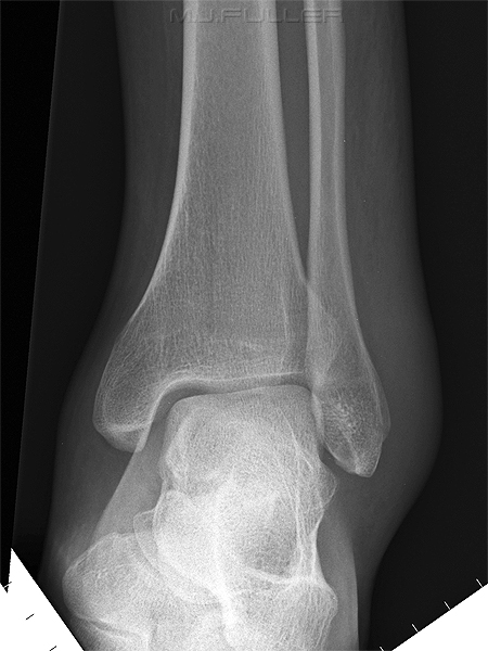
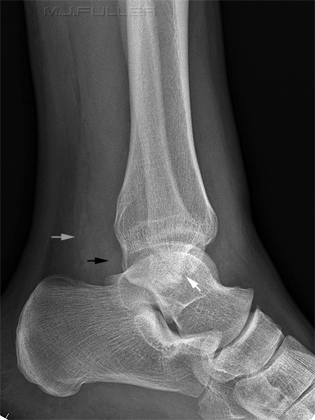
There is significant soft tissue swelling around the ankle, particularly over the lateral malleolus.
No clearly evident fracture.
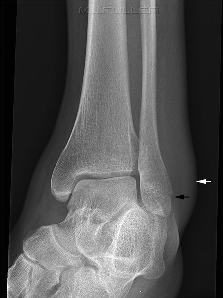
There is further evidence of a distal fibula fracture (black arrow)
Soft tissue swelling again noted (white arrow)
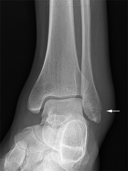
The radiographer repeated the oblique ankle view with slightly different obliquity.
The distal fibula fracture is clearly demonstrated (white arrow)
Discussion
It could be argued that the distal fibula fracture was evident on the initial AP and lateral ankle images. It would also be reasonable to say that the repeated oblique ankle left no doubt as the the presence of the fracture


