Shoulder- SI vs Lateral Scapula

From wikiRadiography
Revision as of 17:39, 11 November 2020 by Travis (talk | contribs) (Created page with "<div class="WPC-editableContent"><b><font size="5">Introduction</font></b><br/><blockquote> Some imaging departments have adopted the lateral scapula view as a routine projec...")
(diff) ← Older revision | Latest revision (diff) | Newer revision → (diff)
Jump to navigation Jump to search
Introduction

Some imaging departments have adopted the lateral scapula view as a routine projection for shoulder trauma radiography. Other institutions strictly forbid the lateral scapula view for shoulder trauma radiography. This page examines three cases which are arguably instructive in this debate.


Case 1. Shoulder Fracture Dislocation
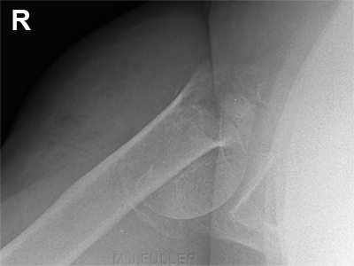
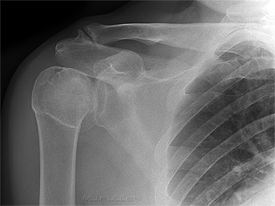
shoulder fracture dislocation This patient presented to the Emergency Department following a fall. The initial AP shoulder image demonstrated an abnormal looking humeral head and neck. The exact nature of the bony deformity was unclear.
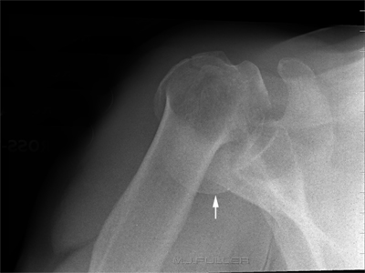
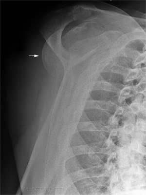
shoulder fracture dislocation The lateral scapula image appearance was unusual, particularly the position of the articular surface of the humeral head (arrowed).
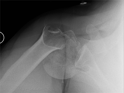
shoulder fracture dislocation The Infero-superior projection (IS) image revealed that the humeral head was dislocated and impacted on the glenoid resulting in a Hill-Sachs impaction fracture.

The patient had an ORIF the following day.


Case 2. Shoulder Fracture Dislocation
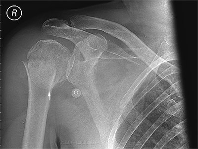
shoulder fracture-dislocation This patient also presented to the Emergency Department following a fall. The initial AP shoulder image revealed a fracture involving the humeral head and neck. One unusual feature of this image was the articular surface of the humeral head appeared to be directed inferiorly (arrow)
shoulder fracture dislocation The infero-superior projection (IS) image revealed that the humeral head was dislocated and impacted on the glenoid resulting in Hill-sachs impaction fracture.

A closed reduction of the shoulder dislocation was attempted under anaesthetic but was unsuccessful. The patient subsequently underwent an operative reduction internal fixation (ORIF).


Case 3. Shoulder Fracture Dislocation?
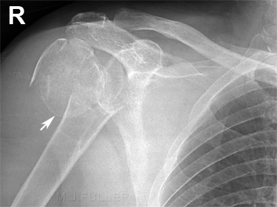
ap shoulder NOH fracture This patient presented to the Emergency Department following a fall. The initial AP shoulder image revealed a fracture involving the humeral head and neck. Not unlike the case above, the articular surface of the humeral head appeared to be orientated inferiorly. The radiographers recognised the importance of establishing whether this was a fracture or a fracture/dislocation. Further view(s) were required
shoulder NOH fracture IS The infero-superior projection (IS) image revealed that the humeral head was not dislocated.

A closed reduction of the shoulder dislocation was performed under anaesthetic to achieve a more satisfactory fracture position. This case demonstrated once again the importance of the IS/SI view of the shoulder in cases of comminuted fracture of the humeral head/neck.


Case 4
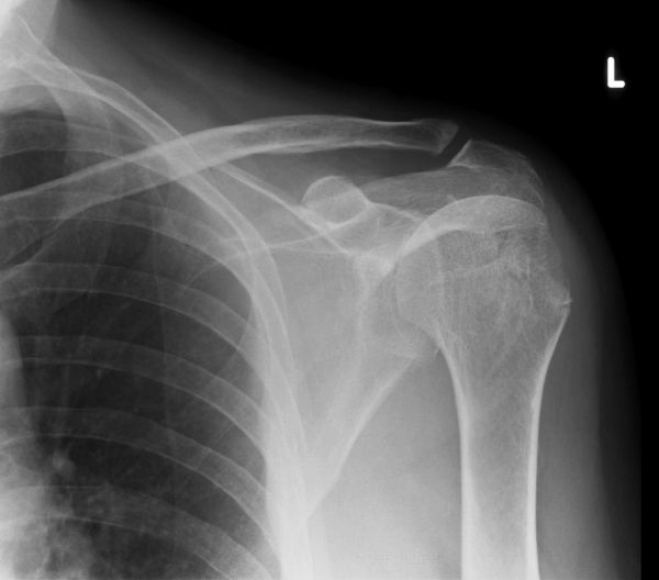
ap shoulder This patient presented for follow-up of a left shoulder dislocation. The AP shoulder image demonstrates fracture(s) involving the greater tuberosity. The alignment of the glenohumeral joint is unclear.
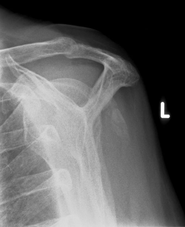
lateral scapula The subacromial projection (approximates lateral scapula projection) demonstrates the fracture fragments. The glenohumeral joint appears to be normally aligned.
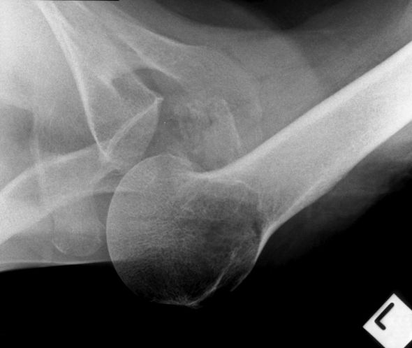
SI shoulderThe SI projection image demonstrates subluxation of the glenohumeral joint.


Comment

Subluxation of the glenohumeral joint (indicating joint laxity) can be missed if the SI projection is not included in the series.


Radiography

It is common to experience difficulty with the IS view in bedbound patients with shoulder fractures. The patients are reluctant to move and the patient's neck sometimes limits medial positioning of the cassette. It can be useful to employ a very long ffd ie instead of placing the X-ray tube beside the patient's torso, increase the ffd such that it is more inferior than the patient's feet (approximately 180cm ffd). Another positioning trick is to place the cassette above the patient's head. Irrespective of the approach that you use, innovation may be required and the image may not be aesthetically pleasing. The lack of image aesthetics should not mitigate against utilising this view (function before form).


Discussion

It might appear by the choice of cases that I have taken a clear position in favour of the IS view over the lateral scapula projection. This is not the case. Both of these cases were drawn from a hospital that does not require the IS/SI view as a routine view in trauma radiography of the shoulder. The routine views adopted by this institution are AP shoulder and lateral scapula. In the first two cases, the radiographer identified the case as a potential fracture-dislocation from the AP and lateral scapula images and performed the SI view to prove the suspicion.

You could safely argue that the institutions that have adopted the IS/SI view as routine in shoulder trauma radiography have taken a safer position. It could also be argued that the institutions that require routine AP shoulder and lateral scapula views, and that have a comprehensive radiography continuing education program, will allow the radiographers to exercise judgment in these types of cases and perform supplementary views as required.

(please contribute your opinion/experiences using the start a new thread link at the bottom of this page)
Relevant WikiRadiography Links


back to the applied radiography home page