Lateral Chest Case 14

From wikiRadiography
Revision as of 17:04, 11 November 2020 by Travis (talk | contribs) (Created page with "<div class="WPC-editableContent"><font size="5"><b>Preamble</b></font><br/> <blockquote>This is the answer page to Case 14 from the page titled What is the Value of the Late...")
(diff) ← Older revision | Latest revision (diff) | Newer revision → (diff)
Jump to navigation Jump to search
Preamble

This is the answer page to Case 14 from the page titled What is the Value of the Lateral Chest Projection?


Other Relevant Wikiradiography Pages


PA Chest

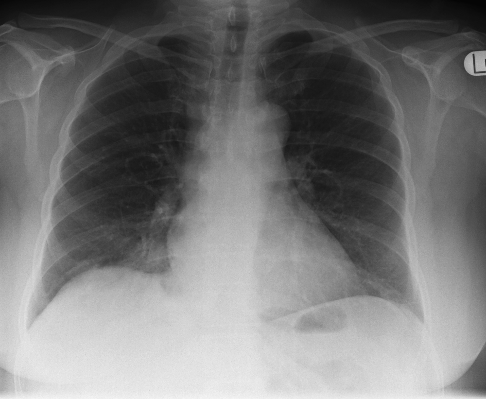
PA chest
This 54 year old female presented to the Emergency Department with a chest infection.
She had a recent history of overseas travel.
There is no clear evidence of lung disease.
The right subdiaphragmatic region was noted to be denser when compared to the left- this can be a very subtle sign of basal lung disease


Lateral
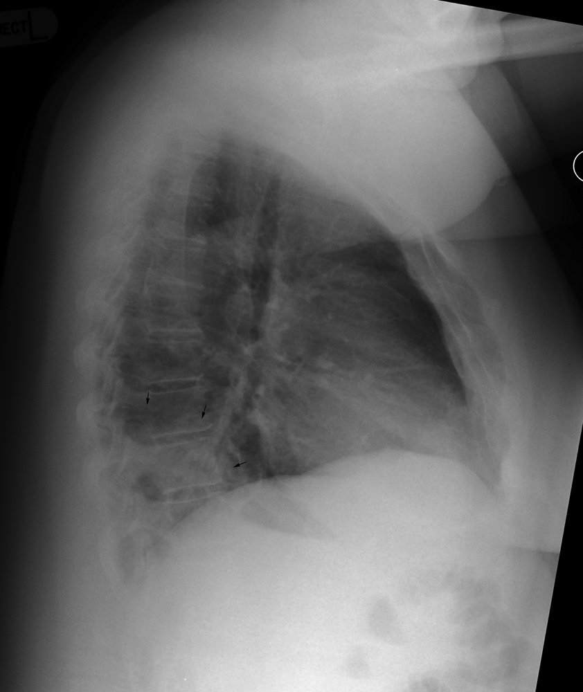
lateral chest
There is abnormal retrocardiac density demonstrated superimposed over the thoracic spine (arrowed)
There is loss of visualisation of the right hemidiaphragm posteriorly (silhouette sign)
This patient is likely to have a pneumonic infection resulting in abnormal alveolar opacity


... back to the wikiRadiography home page
... back to the Applied Radiography home page
... back to the What is the Value of the Lateral Chest Projection? page