Lateral Chest Case 12

From wikiRadiography
Revision as of 17:04, 11 November 2020 by Travis (talk | contribs) (Created page with "<div class="WPC-editableContent"><font size="5"><b>Preamble</b></font><br/> <blockquote>This is the answer page to Case 12 from the page titled What is the Value of the Late...")
(diff) ← Older revision | Latest revision (diff) | Newer revision → (diff)
Jump to navigation Jump to search
Preamble

This is the answer page to Case 12 from the page titled What is the Value of the Lateral Chest Projection?


Other Relevant Wikiradiography Pages
Lung Anatomy


PA Erect
PA chest
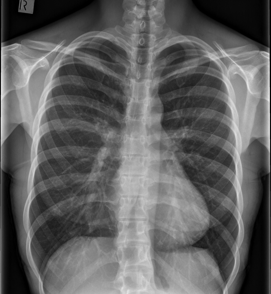
This 24 year old female was referred for chest radiography with a possible axillary DVT.
The chest radiography was preformed to assess possible cervical ribs.
Imaging was performed with a DR system
There is loss of visualisation of the right heart border
There is a mild scoliosis concave to the left
11 posterior ribs are seen above the diaphragm indicating overexpansion of the lungs
Note the upward curvature of the posterior ribs that can be associated with pectus excavatum


Lateral
lateral chest
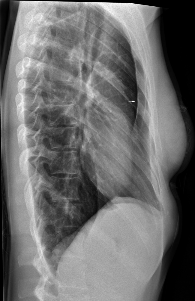
There is pectus excavatum (arrowed) which is associated with loss of visualisation of the right heart border
Pectus excavatum refers to the depressed inferior sternum.
This condition can leave insufficient space for the heart between the sternum and thoracic vertebral bodies causing the heart to displace to the left.
It is this displacement to the left that results in the failure of visualisation of the right heart border


... back to the wikiRadiography home page
... back to the Applied Radiography home page
... back to the What is the Value of the Lateral Chest Projection? page