Imaging Tibial Plateau Fractures
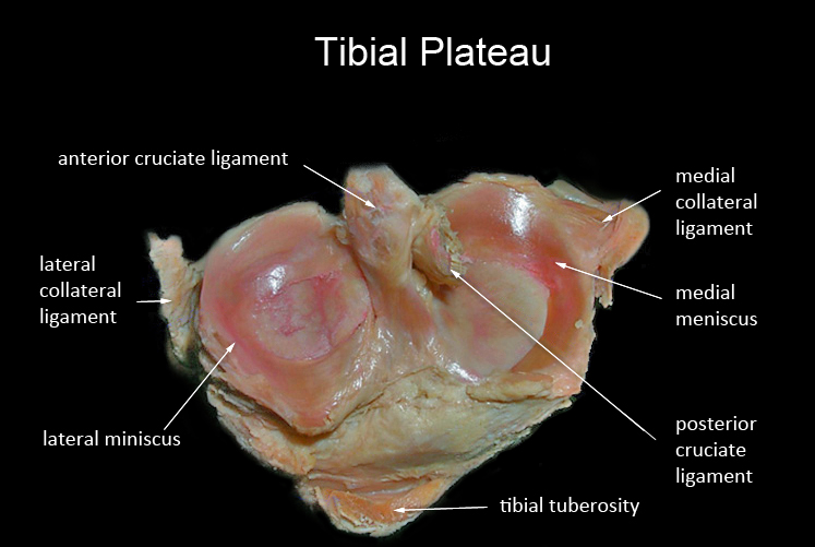
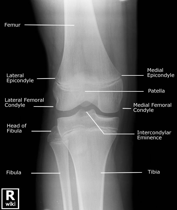
AnatomyA full and adequate radiographic demonstration of a tibial plateau fracture is a hallmark of an accomplished trauma radiographer. A clinical approach to radiography with careful consideration of radiographic soft tissue signs is essential. This page considers all aspects of the radiography of tibial plateau fractures.
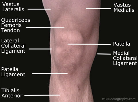
based on<a class="external" href="http://www.ithaca.edu/faculty/lahr/LE2000/LE_knee.html" rel="nofollow" target="_blank"> Ithaca College</a><a class="external" href="http://www.ithaca.edu/faculty/lahr/LE2000/LE_knee.html" rel="nofollow" target="_blank"> Department of Physical Therapy</a><a class="external" href="http://www.ithaca.edu/faculty/lahr/LE2000/LE_knee.html" rel="nofollow" target="_blank"> Human Anatomy Review Site, http://www.ithaca.edu/faculty/lahr/LE2000/LE_knee.html</a>The medial and lateral miniscii provide a cushion for the weightbearing forces transmitted from the condyles of the femur. In cases of suspected tibial plateau fracture, examination of the patient's knee can provide evidence of the underlying injury. In particular, it is worthwhile considering whether the patient has a knee effusion and whether there is evidence of varus or valgus deformity at the knee joint.
Notes on Tibial Plateau Fractures
- condition of fibula, whether fractured or intact, determines angulatory behavior of these fractures under weight bearing and functional conditions
- isolated fractures of the lateral plateau with intact fibula do not tend to collapse further because of the support of the fibula- by contrast, fractures of both lateral tibial plateau and fibula have tendency to collapse into valgus because of the loss of support of the fibula<a class="external" href="http://www.wheelessonline.com/ortho/role_of_fibula_in_tibial_plateau_fractures" rel="nofollow" target="_blank">http://www.wheelessonline.com/ortho/role_of_fibula_in_tibial_plateau_fractures</a>
Case 1
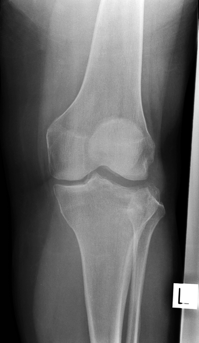
This 65 year old lady presented to the Emergency Department following a fall from a ladder. The patient was examined and referred for left knee radiography. The patient thought that she had 'broken a bone in her knee'.
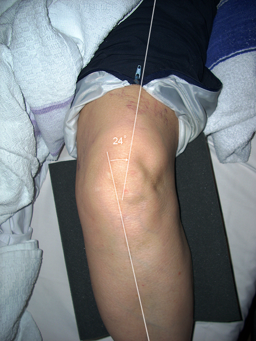
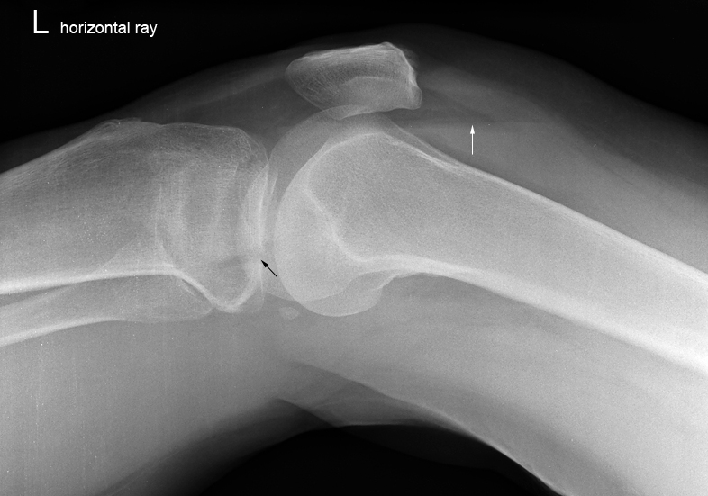
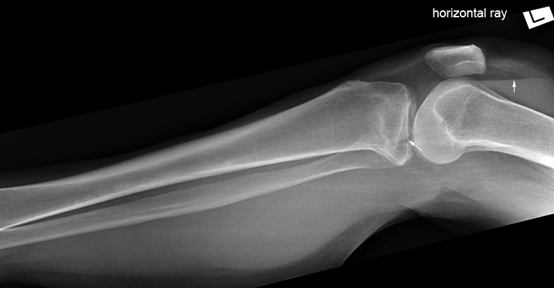
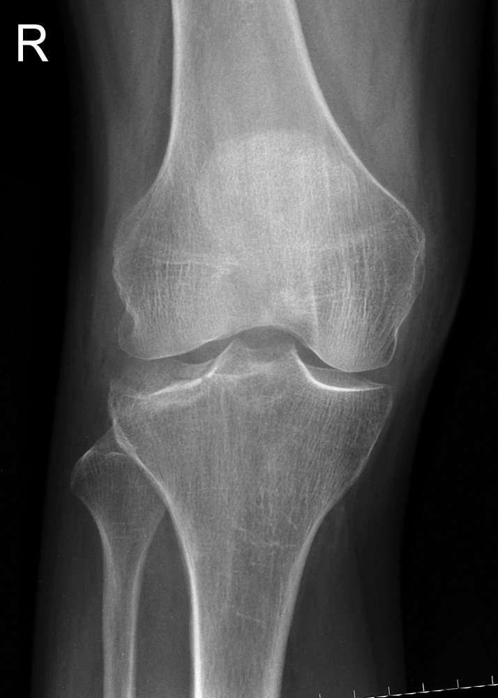
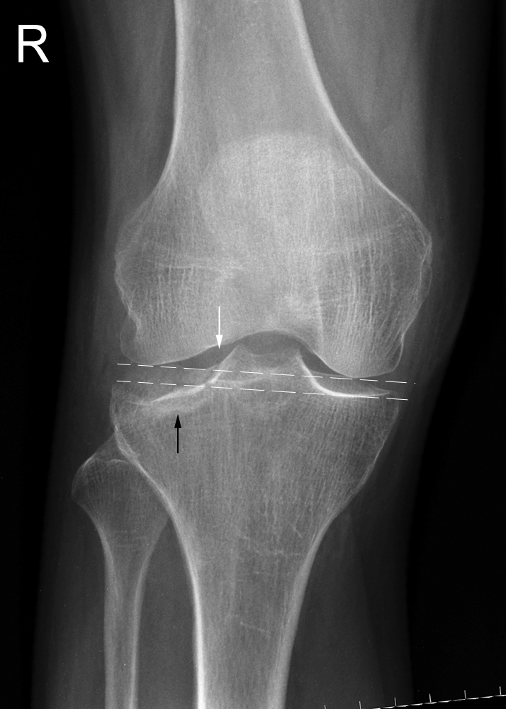
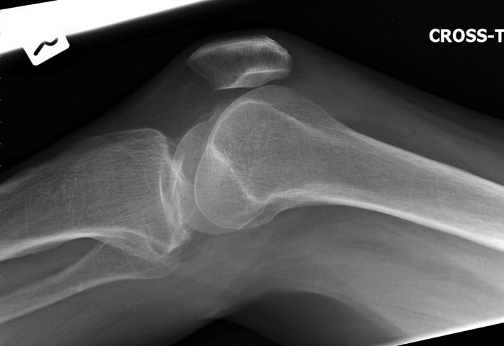
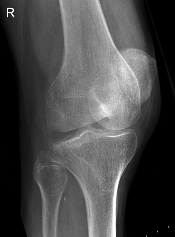
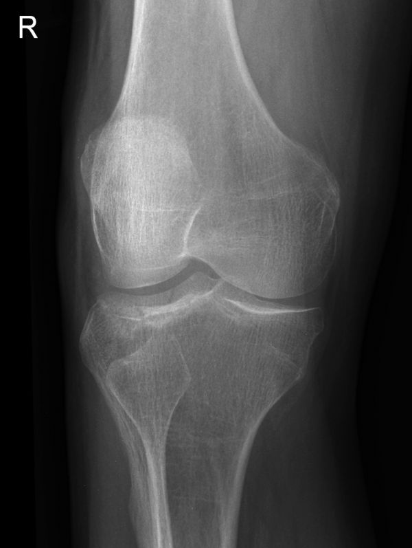
The radiographer noted a swollen knee and fluid in the suprapatellar pouch suggesting a knee effusion. In addition, there appeared to be a valgus deformity at the knee joint (normal Q angle 10 - 22 degrees)The AP knee projection image suggested a depressed tibial plateau fracture laterally. The horizontal ray lateral knee image demonstrated a lipohaemarthrosis (white arrow) and there was a suggestion of a discontinuity of the cortical contour of the tibial plateau (black arrow).
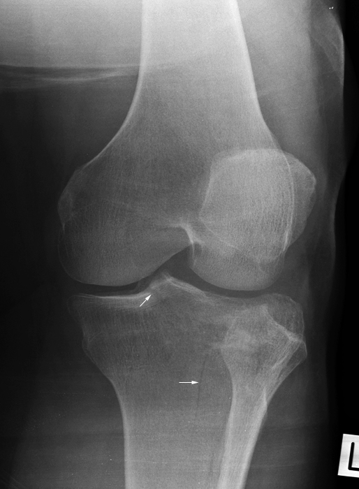
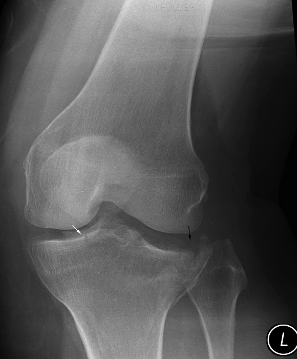
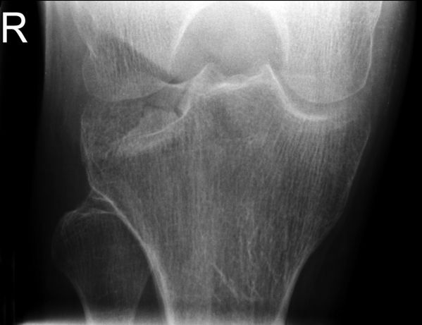
The radiographer suspected a tibial plateau fracture and proceeded to perform oblique knee projections.The patient was in too much pain (despite pain relief) to rotate her leg for oblique knee projections. The radiographer performed the oblique projections using tube angulation rather than rotating the patient's knee internally and externally.
(left) This is the equivalent of an external rotation projection of the knee. There is evidence of a fracture at the base of the tibial spines and a vertical fracture of the metaphysis of the proximal tibia (bottom arrow). Note the disruption of the normal trabecular pattern of the lateral tibial metaphysis and the associated sclerotic bands.This is the equivalent of an external rotation projection of the knee. The tibial spine fracture is again evident (white arrow) and the depressed fracture of the lateral articular surface of the tibia is well demonstrated. The radiographer performed additional views of the patient's lower leg to establish whether the fracture extended further down the tibia. The now sharply delineated lipohaemarthrosis (white arrow) may reflect "snowglobe effect".
Case 1 Notes
- The recognition of the knee lipohaemarthrosis by the radiographer and the supplementary oblique knee views should be considered to be an appropriate minimum standard of radiography in this case
- If the orthopaedic surgeon was available (not so in this case) and knee CT was planned, the oblique projections may not have been warranted.
Case 2
