Imaging Shoulder Dislocations
Introduction
Dislocations of the glenohumeral joint are commonly seen in Emergency Departments. This page considers all aspects of radiography of glenohumeral joint dislocations
Anatomy
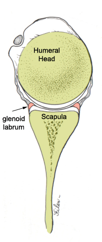
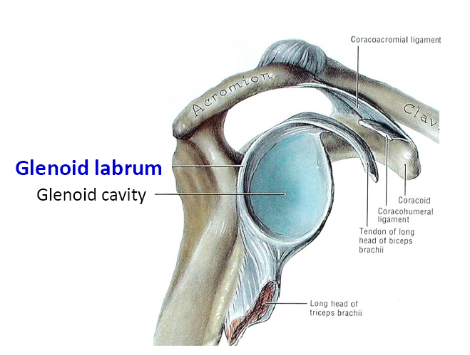
These videos provide information on the bony and soft tissue anatomy of the shoulder
<embed allowfullscreen="true" height="350" src="http://widget.wetpaintserv.us/wiki/wikiradiography/widget/youtubevideo/555b6a27dc7a09ec6b0cf1e2fc371b5ec06321ef" type="application/x-shockwave-flash" width="425" wmode="transparent"/> <embed allowfullscreen="true" height="350" src="http://widget.wetpaintserv.us/wiki/wikiradiography/widget/youtubevideo/2bfdb94ac3b82565b8f0f6184d05959b093b29dc" type="application/x-shockwave-flash" width="425" wmode="transparent"/> <embed allowfullscreen="true" height="350" src="http://widget.wetpaintserv.us/wiki/wikiradiography/widget/youtubevideo/088c607c7711350d0d6e8b973b64d988c14460e0" type="application/x-shockwave-flash" width="425" wmode="transparent"/> <embed allowfullscreen="true" height="350" src="http://widget.wetpaintserv.us/wiki/wikiradiography/widget/youtubevideo/a228652a643fb0f13e9f8f631756121c39e23b29" type="application/x-shockwave-flash" width="425" wmode="transparent"/> <embed allowfullscreen="true" height="350" src="http://widget.wetpaintserv.us/wiki/wikiradiography/widget/youtubevideo/27a864b1476e85345ce0357a63679a1906b3d24e" type="application/x-shockwave-flash" width="425" wmode="transparent"/> <embed allowfullscreen="true" height="350" src="http://widget.wetpaintserv.us/wiki/wikiradiography/widget/youtubevideo/c7a2ac1393c0aee9ae4175d33c299d5e7d80afbe" type="application/x-shockwave-flash" width="425" wmode="transparent"/>
Golf Ball on a Tee Analogy
source: unknown<a class="external" href="http://www.manywallpapers.com/pop_preview.html/-/p/golf-ball/id/71502" rel="nofollow" target="_blank">http://www.manywallpapers.com/pop_preview.html/-/p/golf-ball/id/71502</a>
Clinical Presentation
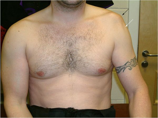
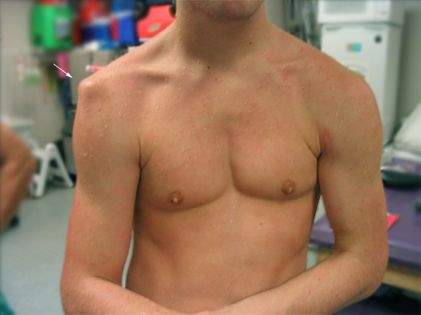
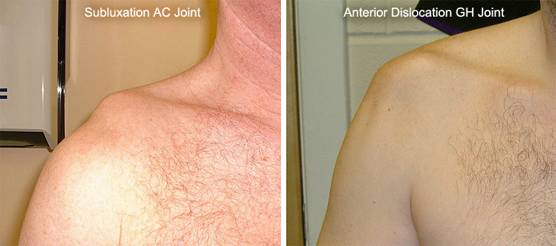
source: unknownThe anterior dislocation often presents with a highly characteristic appearance. The shoulder will be 'squared off' (black arrow) and a skin depression will be evident (white arrow).
-loss of deltoid muscle contour compared to contra-lateral side
-humeral head is sometimes palpable anteriorly beneath the coracoid
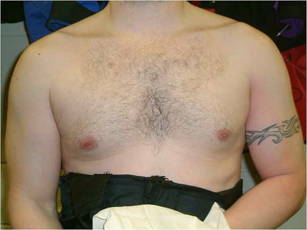
source: unknownFollowing a relocation procedure, the normal shoulder contour is re-established.
Note the red skin discolouration- this is caused by the considerable force exerted by the person who relocated the dislocated glenohumeral joint in this male with well developed musculature
<a class="external" href="http://whs.wsd.wednet.edu/Faculty/Blair/sportsmed/shdlrdisl.JPG" rel="nofollow" target="_blank">http://whs.wsd.wednet.edu/Faculty/Blair/sportsmed/shdlrdisl.JPG</a>Dislocated right shoulder with typical 'squaring off'.
Radiographic Appearances of Shoulder Dislocations
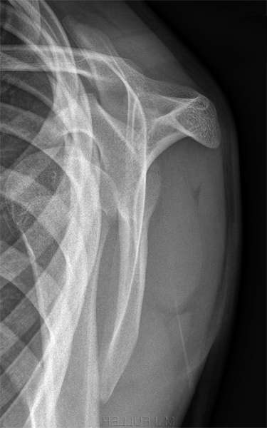
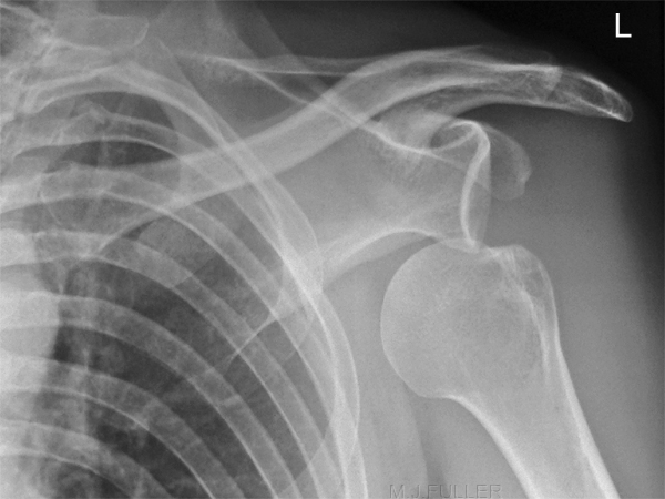
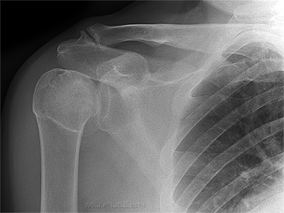
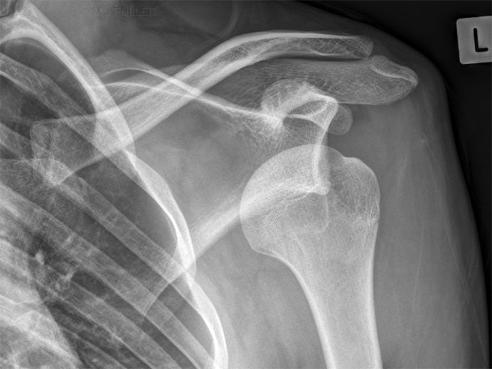
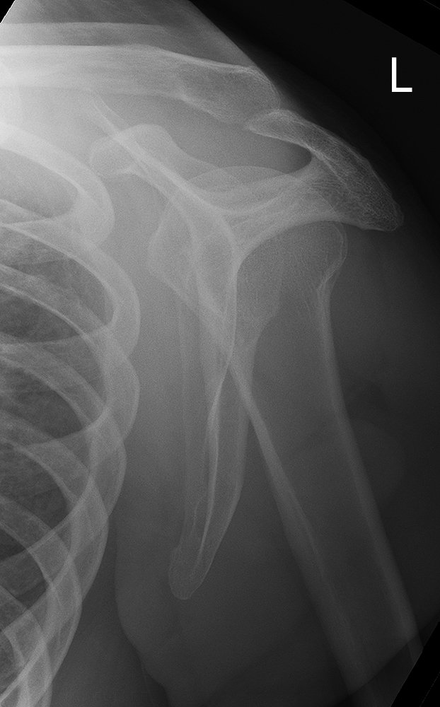
Normal Shoulder
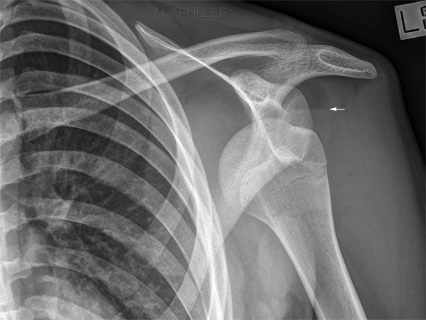
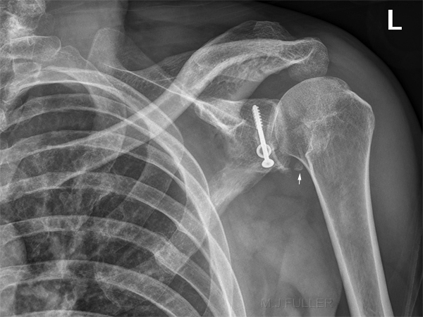
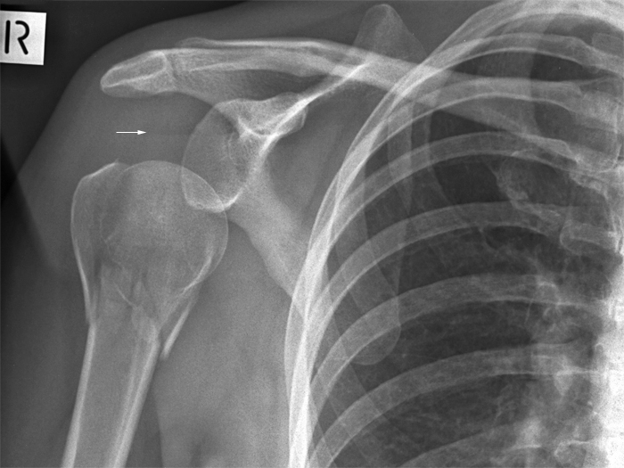
Anterior Shoulder Dislocation
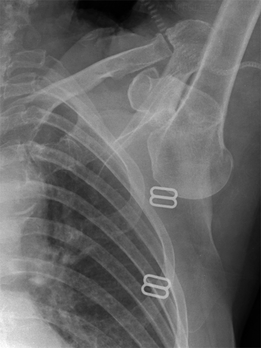
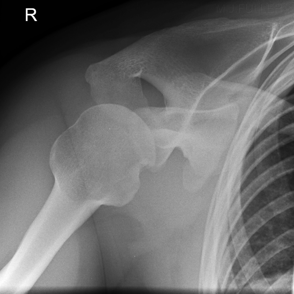
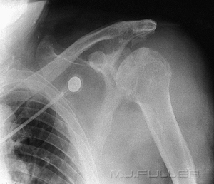
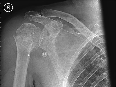
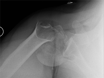
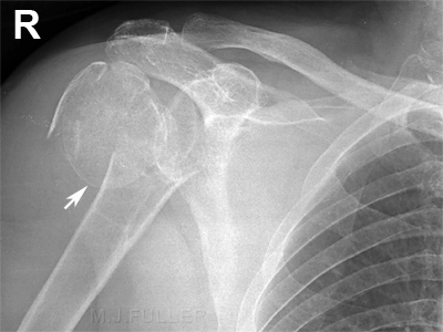
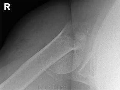
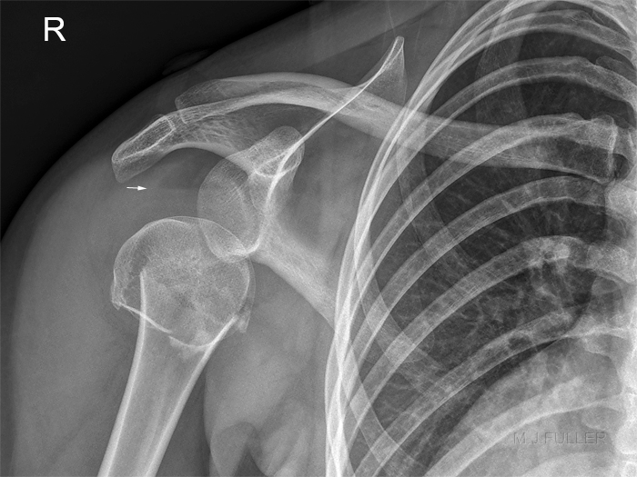
This patient has an anterior shoulder dislocation. The arrowed horizontal line is a lipohaemarthrosis within the shoulder joint. The humeral head is located anterior to the glenoid.
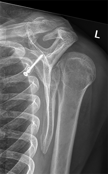
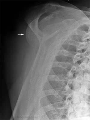
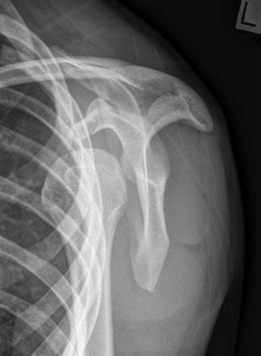
Posterior Shoulder Dislocation
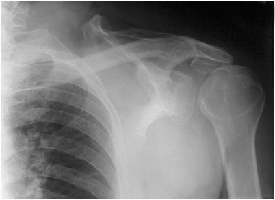
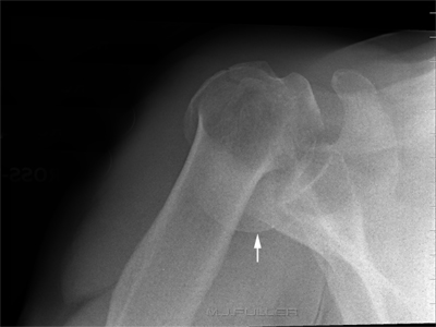
Inferior Shoulder Dislocation 1
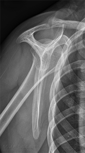
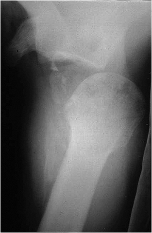
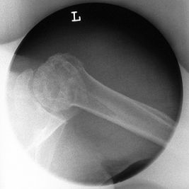
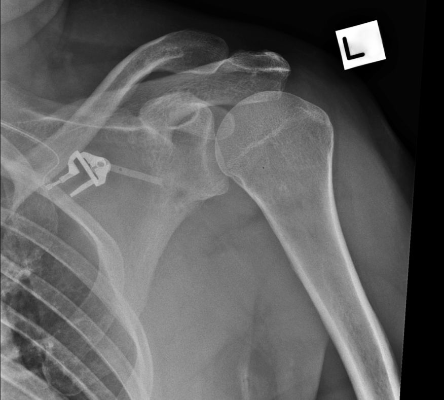
This patient has an inferior dislocation of the glenohumeral joint (Luxatio erecta). The SI view image shows the humeral head to be dislocated slightly anteriorly.
Inferior Shoulder Dislocation 2 Luxatio erecta
This patient has an inferior dislocation of the glenohumeral joint which is referred to as Luxatio erecta. Inferior shoulder dislocation (1-2%)
- Luxatio erecta ─ uncommon form of shoulder dislocation
- Extremity held over head in fixed position with elbow flexed
- Mechanism
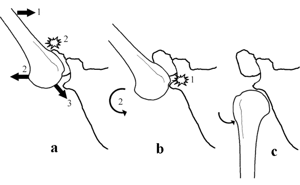
· Severe hyperabduction of arm resulting in impingement of humeral head against acromion
· Humeral articular surface faces inferiorly
<a class="external" href="http://www.learningradiology.com/archives06/COW+200-Luxatio+erecta/luxatioerectacorrect.htm" rel="nofollow" target="_blank">quoted from: www.learning radiology.com</a>This patient has an inferior dislocation of the glenohumeral joint.
Source:
<a class="external" href="http://www.josr-online.com/content/4/1/40" rel="nofollow" target="_blank">S Saseendar, Dinesh K Agarwal, Dilip K Patro and Jagdish Menon
Unusual inferior dislocation of shoulder: reduction by two-step maneuver: a case report
Journal of Orthopaedic Surgery and Research 2009, 4:40</a>
"Inferior dislocation of the shoulder (luxatio erecta) is rare accounting for an estimated 0.5% of shoulder dislocations. First described by Middeldorpf and Scharm in 1859, it occurs when the shoulder is forced into hyperabduction with the proximal humerus being levered over the acromion process. It may also result from direct axial loading on the fully abducted arm. The humerus is locked in a position somewhere between 110 and 160 degree of adduction. Severe soft tissue injury or fractures about the proximal humerus occur with this dislocation. It is estimated that 60% of these patients have some neurological deficit (brachial plexus injury) of the upper limb prior to reduction. Axillary artery injury is another possible serious complications. Associated bony injuries include fractures of the greater tuberosity, acromion, clavicle, coracoid process, and glenoid rim. Long-term complications include adhesive capsulitis and recurrent subluxations or dislocations. Radiological investigation will show the shaft of the humerus lying parallel to the spine of the scapula and the articular surface of the humeral head directed inferiorly without contact with the glenoid."
quoted from <a class="external" href="http://www.mskcases.com/index.php?module=article&view=120" rel="nofollow" target="_blank">http://www.mskcases.com</a>
Fracture Dislocations of the Shoulder
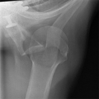
In a shoulder fracture/dislocation, the associated humeral fracture is almost always displaced
Anterior dislocations are associated with fractures of the greater tuberosity and posterior dislocations are associated with fractures of the lesser tuberosity
Modified Technique for Trauma Patients 1
Modified Technique for Trauma Patients 2
Modified Technique for Trauma Patients 3
Can the Lateral Scapula Projection Reliably Demonstrate Shoulder Dislocation?
My department has utilised the lateral scapula projection as the view of choice for the demonstration of shoulder dislocations for the last 30 years. There are departments that strictly forbid the lateral scapula view for assessment of shoulder dislocation. They can't both be correct ... or can they?
The Counter-Argument
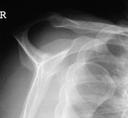
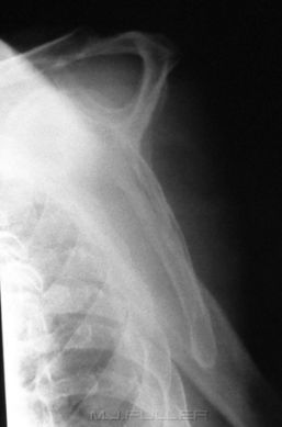
The counter argument is that there are several conditions where the results can be equivocal. Amongst these conditions are the pseudosubluxation and the posterior dislocation.
Discussion
The limitations of the lateral scapula view can be overcome with radiographer training. Radiographers learn to identify cases where the lateral scapula view should be supplemented with views such as the IS/SI view.
In one study, it was found that " .... the axillary view and scapular "Y" view visualized associated pathology equally well" Silfverskiold JP. Straehley DJ. Jones WW. Orthopedics - pcm. [JC:pcm] 13(1):63-9, 1990 Jan quoted in <a class="external" href="http://www.wheelessonline.com/ortho/roentgenographic_evaluation_of_suspected_shoulder_dislocation_a" rel="nofollow" target="_blank">Wheeless on Line</a>
Bilateral Shoulder Dislocations
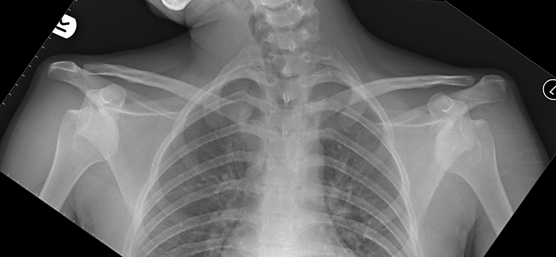
Pseudosubluxation
Lateral Scapula vs SI Shoulder
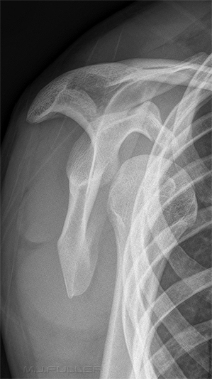
Some imaging departments have adopted the lateral scapula view as a routine projection for shoulder trauma radiography. Other institutions strictly forbid the lateral scapula view for shoulder trauma radiography. This page examines three cases which are arguably instructive in this debate.
Case 1. Shoulder Fracture Dislocation
Case 2. Shoulder Fracture Dislocation
Case 3. Shoulder Fracture Dislocation?
Radiography
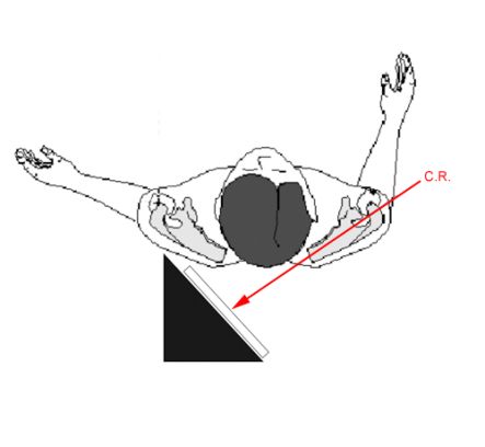
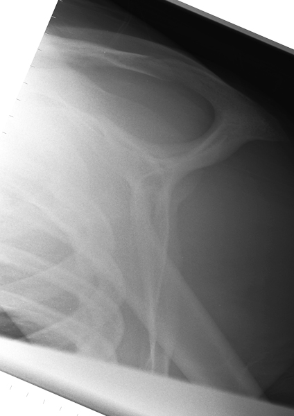
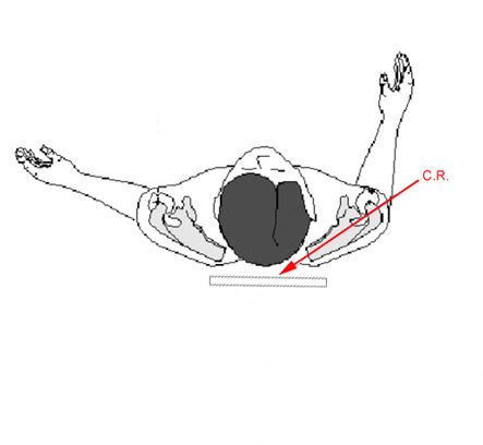
It is common to experience difficulty with the IS view in bedbound patients with shoulder fractures. The patients are reluctant to move and the patient's neck sometimes limits medial positioning of the cassette. It can be useful to employ a very long ffd ie instead of placing the X-ray tube beside the patient's torso, increase the ffd/SID such that it is more inferior than the patient's feet (approximately 180cm ffd). Another positioning technique is to place the cassette above the patient's head. Irrespective of the approach that you use, innovation may be required and the image may not be aesthetically pleasing. The lack of image aesthetics should not mitigate against utilising this view (function before form).
DiscussionIt might appear by the choice of cases that I have taken a clear position in favour of the IS view over the lateral scapula projection. This is not the case. Both of these cases were drawn from a hospital that does not require the IS/SI view as a routine view in trauma radiography of the shoulder. The routine views adopted by this institution are AP shoulder and lateral scapula. In the first two cases, the radiographer identified the case as a potential fracture-dislocation from the AP and lateral scapula images and performed the SI view to prove the suspicion.
You could safely argue that the institutions that have adopted the IS/SI view as routine in shoulder trauma radiography have taken a safer position. It could also be argued that the institutions that require routine AP shoulder and lateral scapula views, and that have a comprehensive radiography continuing education program, will allow the radiographers to exercise judgment in these types of cases and perform supplementary views as required.
Lipohaemarthrosis
Case 1
Case Studies
Case 2
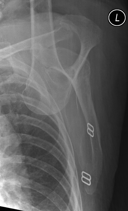
This 51 year old male presented to the Emergency Department by ambulance after a RTA/MVA. He was examined in the resus room and a variety of radiographic examinations were requested including left shoulder. The patient was experiencing considerable pain in the left hemithorax and sternum and could therefore not be moved from the supine position.
The patient's shoulder was in internal rotation. An AP shoulder projection was achieved with the patient ina supine position with tube angulation towards the left shoulder (35 - 45 degrees).
A <a class="external" href="http://www.carestreamhealth.com/PublicContent.aspx?langType=1033&id=448841" rel="nofollow" target="_blank">CARESTREAM DRX-1</a> System was utilized with no grid. The patient tolerated the DRX cassette directly under the shoulder.Using the DRX cassette the cassette could be left under the patient fro the lateral scapula projection. The beam was angled 35 - 45 degrees towards the right shoulder to good effect. The resultant image is distorted (elongated) but acceptable under the circumstances.
Relevant WikiRadiography Links
